Download
1 / 61
640 likes | 966 Views
Minimally Invasive Cancer Therapies in Interventional Radiology. Chief, Vascular and Interventional Radiology Lancaster Radiology Associates Co-Director, Interventional Vascular Unit. Objectives. 1- Identify currently available IR procedures related to cancer care at LGH

E N D
Minimally Invasive Cancer Therapies in Interventional Radiology Chief, Vascular and Interventional Radiology Lancaster Radiology Associates Co-Director, Interventional Vascular Unit
Objectives • 1- Identify currently available IR procedures related to cancer care at LGH • 2- Enhance medical staff knowledge of such procedures • 3- Discuss current IR cancer treatments
Palliative and curative therapies • Diagnosis • Lung • Genitourinary • Gastrointestinal
DIAGNOSIS through Image-Guided Biopsies • Often one of the initial procedures used to obtain a tissue diagnosis • Multiple modalities including Computed Tomography, Ultrasound, and Fluoroscopy • Alone or in combination • Often correlate with PET scan to identify “active” sites
Biopsy Technique • Often coaxial with “outer” introducer needle and “inner” biopsy needle • Need a “window”; Want to obtain an adequate tissue sample for diagnosis but need to utilize a safe approach • May use conscious sedation along with local anesthesia
Ultrasound biopsies • Require hand-eye coordination • May be used for random sampling, i.e. for gross liver biopsy • For focal lesions, often in difficult to access locations, if poorly seen on CT scan, or if lesion is “mobile”
X-ray guided biopsy • Especially useful when patient positioning is limited; can rotate and angle the tube to obtain an approach for lesion access • Advantage of real time imaging
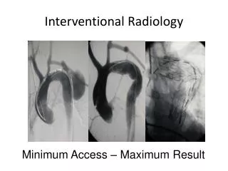
Rotational angiography and Xper CT Technology in new Philips angio equipment that combines CT and 3D-imaging. Enhances IR procedures by allowing you to import previous MRI or CT data and fuse it with angiographic studies. Allows the interventionalist to use fluoroscopy and apply it to a CT image for challenging access.
Lung Palliative Tunneled pleural catheters Thermal ablation of destructive chest wall lesions Curative RFA of unresectable lung cancers or lung metastases
RFA lung cancer • Early NSCLC or metastases in those deemed NOT to be surgical candidates • Could have a poor functional status, abnormal PFTs’, Octogenarians?etc. • Relapse in Radiation field • Painful bone metastasis • Chest wall invasion
Lung Cancer survival • If untreated, median survival 9-12 months. • Surgical resection 5 year 60-70% • RFA or Radiation 5 year 30-50% • RFA 1 yr: 83-90%; 2 year 48-83%
LGH statistics • 20 tumors treated with RFA; 16 patients. • Treatment goals met in 15/16 patients. All but one patient was treated for cure. • 4/16 patients required an additional ablation. • Stable or without recurrence for up to 26 months. • 1 unrelated death two days after treatment. Cardiac arrest.
Genitourinary (GU) • Palliative • Percutanousnephrostomy • Dialysis catheters • Fistula or hemodialysis access maintenance • Curative • Thermal ablation of renal cell cancer
GU procedures • Percutaneous access to the collecting system for benign or malignant obstructions, stone disease, or urosepsis • Can place internal double J ureteral stents from percutaneous access • Can provide access for future stone removal and/or manipulation
Cryoablation • Argon gas for freezing; Helium for thawing. • Multiple probes; RFA just a single probe. • Less risk of damage to collecting system. • Greater risk of bleeding compared with RFA (coagulative necrosis). • -20 to -40 degrees Celsius. Cell death. • Can better identify treated zone.
Survival • Stage I RCC- surgery with partial nephrectomy or nephrectomy 80+% 5 year survival • Difficult to do much better for early disease • Stage I RCC treated with RFA for 3 cm tumors or smaller 94% 2 year survival. Decreased survival as tumor size increases beyond 3 cm.
Is RCC Cryoablation Effective? 19 months 26 months 24 months • 1Littrup, J VascIntervRadiol 2007; Atwell, J Urol 2010; • Rodriguez, CardiovascIntervRadiol 2011
LGH statistics • 7 tumors treated • 6/7 Renal cell cancer. 1/7 benign oncocytoma. • 6/7 no signs of recurrence. 1/7 partially treated and opted for surveillance.
Gastrointestinal (GI) • Palliative • Peritoneal catheters • Gastric tubes • Cholecystostomy drains • Biliary stents • Locoregional control • Catheter-based embolization • Percutaneous thermal ablation
Image-Guided Therapy for Hepatic Malignancies Liver Dominant Unresectable
Definitions • Liver-dominant neoplasm: malignancy in which the hepatic component is the only site of disease or the dominant site most likely to lead to patient morbidity or mortality
What’s so good about embolization or chemoembolization? • Minimally-invasive loco-regional treatment • Spares the patient the morbidity of surgery, radiation, or systemic therapy • Achieves tumor necrosis • Increases drug concentration delivered and dwell time of agent(s) • Decreases systemic toxicity
Definitions • Embolization: refers to blocking arteries by particles alone • Oily Chemoembolization: infusion of chemotherapeutic agents with Ethiodized oil followed by embolic agents • Drug-eluting beads: chemoembolization with calibrated microspheres that release drug over time
Definitions • Tumor Ablation: direct application of thermal or chemical therapies to tumor(s) to eradicate or substantially destroy it • Chemical: ethanol or acetic acid • Thermal: application of energy to cause tumor necrosis. Examples include radiofrequency ablation (RFA), microwave, cryotherapy, high-intensity focused ultrasound (HIFU)
Why consider tumor ablation? • Patients are living longer and presenting later in life with cancer. • Co-morbid conditions are a major factor in considering patients for surgical resection. • Minimally invasive therapies are in demand. • Tumor ablation offers a chance for cure without surgery. • Important psychological benefits to patients instead of just waiting and seeing what happens.