Download
1 / 38
430 likes | 515 Views
Interventional Radiology in Pediatric Trauma . C. Moorthy, MD. Disclosures . No disclosures No vender specific equipment endorsed. Wisdom. Kids are not little adults!—except maybe in interventional techniques? Smaller catheters and wires—that’s it?. General Indications.

E N D
Interventional Radiology in Pediatric Trauma C. Moorthy, MD
Disclosures • No disclosures • No vender specific equipment endorsed
Wisdom • Kids are not little adults!—except maybe in interventional techniques? • Smaller catheters and wires—that’s it?
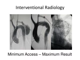
General Indications • Minimally invasive control of bleeding or re-establishment of flow • Percutaneous stenting and drainage to tx. Disruptions of urinary and biliary systems • Tx delayed complications of trauma-embolization of arterial pseudoaneurysms and pulmonary embolism prophylaxis
Statistics • Trauma leading cause of mortality in kids older than 1 yr • 2/3 of trauma related mortality is MVC, followed by homicide, suicide and drowning • Non-fatal trauma mostly falls • ATV, farm injuries, sports, pedestrian and bicycle accidents, birth trauma, iatrogenic, NAT (child abuse)
Trauma physiology/management • Initial sign of shock are subtle and can include lethargy, decreased capillary refill and tachycardia • Increased physiological reserve, so hypotension is a late and ominous sign • If hypotensive (<70mm Hg + 2 x age in years), then pt. has lost approx. 40% BV
Physiology • Primarily dealing with hemorrhagic shock—worsened by the triad of hypothermia, coagulopathy, and acidosis • Decision made by trauma resuscitation team as to initial staged control of bleeding and frank contamination, followed by definitive repair—not to worsen cycle
MDCT • Primary role of MDCT—fast, accurate, and safe (radiation exposure) • Technology driven-number of detectors and radiation sources with protocols configured to provide optimal IV contrast enhancement (dilution and timing) • Almost no role for diagnostic angiography
IR technique-considerations • Embolize as distally as possible to decrease tissue necrosis and lactic acidosis • Pt. weight—determines contrast dosing, fluid (flushes), meds • Contrast 3-5 cc/kg • Radiation dose-Step Lightly campaign. Extension of Image Gently. (SPIR)
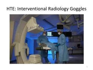
IR-technical considerations • Room temperature 27*C (80*F), blankets, bear hugger • Heat fluids • Vascular access difficult-smaller vessels prone to spasm and thrombosis • Ultrasound guidance standard-high frequency transducers
IR-technical considerations • Infants < 10kg -> 3F sheath and catherters/microcatheters • Child>10kg-> 4F systems, .038 lumen size to accept microcatheter • Use neurovascular and coronary devices • Embolization-metallic coils, gelofam pledgets/slurry, PVA/synthetic particles
IR- technical considerations • Pseudoaneurysms and AV fistulas-soft detachable coils, thrombin injection (US guidance), stent grafts across neck or mouth • Long term outcomes of stent grafts in kids unknown->pseudointimal hyperplasia, and vessel growth. Same problem with surgical grafts • ?biodegradable stent grafts
CNS • Acceleration/decelleration or penetrating • Injury at transition from non-fixed to fixed-skull base and tentorium • Basal skull fxs-CC fistula • Direct penetrating injury ->pseudoaneurysm or traumatic fistula • Imaging shows strokes in vascular pattern
CNS • CTA and MRA techniques better-but go to cath if imaging negative and suspicion high • CC fistula-> occlude fistula • Dissection-> anti-coagulation unless contraindicated. Risk of delayed pseudoaneurysm. F/U CTA or MRA
Thorax • More compliant due to elasticity of ribs, unossified CC junctions, and flexible ligamentous attachments • Increased transmission of force to internal structures->laceration, contusion, ptx, hemothorax • IR involved in delayed tx of loculated collections/thrombolytics
Thorax • PA branch pseudoaneurysm ->coil embolization • Systemic arteries- bronchial, intercostal-> coil embo. ( watch for spinal feeders) • Aorta injury uncommon- no steering wheel, pre-hospital mortality 85%, 50% if those who reach ED dies in 48 hrs
Thorax-aorta • Descending thoracic aorta relatively fixed compared to heart/arch—due to intercostals, ligamentum arteriosum and pleura • Leads to shearing injury at isthmus-intimal, mural, complete • Beware ductus bumpMDCT
Liver • Dual blood supply (80% portal, 20 % arterial) protects from ischemia • Arterial embolization safe is portal vein patent. If portal vein occluded then arterial embolization carries risk of necrosis and/or biliary strictures • Necessitates modified technique
Liver • Non-operative management successful in upto 98% • Hepatic arterial bleeding -> active extravasation, AV fistula, pseudoaneurysm, and arteriobiliary fistula • Can be delayed presentation-jaundice, hematemesis or melena
Liver • As selective as possible • Embolize distal and proximal to pseudoaneurysm, if possible • Direct thrombin injection with US guidance can be done in ICU • Avoid cystic artery occlusion • Traumatic biloma/leak->drain. Combo procedures with GI
Spleen • Most commonly non-operative management • Risk of lowered immunity to encapsulated bacteria and protozoa post splenectomy • So embolize as distal as possible • Unstable or significant capsular breach-can embolize proximally. • Collateral supply from short gastric and pancreatic branches prevent infarction • Prophylactic vaccination
GU system • Renal injury more common than in adults due to relative larger size and mobility • Conservative tx. Mostly • When indicated, intervention is for active extravasation or MRA occlusion • Most common is iatrogenic trauma from biopsy resulting in AV fistulahematuria
GU system • Post biopsy AV fistula -> embolize both afferent and efferent arteries. Pseudoaneurysm-> can inject thrombin directly • Direct perineal straddle injury pudendal artery disruption if connection to corpora then high flow priapism. Embolize with temporary agents • If connection to urethral tear, then presents as hematuria
Pelvis • Uncontrolled pelvic bleeding post-trauma has high mortality • Difficult surgical retroperitoneal exploration which can release tamponade • 90% success from transcatheter embo. • Standard sites—superior gluteal, lateral sacral, internal pudendal and obturator branchesbased on CT
IVC filters • Most common after trauma when contraindication to anticoagulation • Easier now because of removable filters • Evidence that filters can be placed in IVC less than 10 mm without increased risk of caval thrombosis