Download
1 / 27
290 likes | 582 Views
“Iron - Avoiding deficiency” Bronwyn Williams Haematologist – HSSA / RCH. Iron -. Points for Discussion. Iron metabolism and its regulation Prevalence and causes of iron deficiency Diagnostic workup /differential diagnosis Treatment of IDA.

E N D
“Iron - Avoiding deficiency” Bronwyn Williams Haematologist – HSSA / RCH
Iron - Points for Discussion Iron metabolism and its regulation Prevalence and causes of iron deficiency Diagnostic workup /differential diagnosis Treatment of IDA
Metabolic functions of iron – a transition metal Haem iron compounds cytochrome a,b,c (oxidative energy) cytochrome P450 (drug metabolism) catalase, peroxidase (ROS protection) haemoglobin, myoglobin Non-haem iron compounds NAD dehydrogenase (mitochondrial respiration) succinate dehydrogenase xanthine oxidase (nucleotide catabolism) ribonucleotide reductase (nucleotide synthesis) Fairbanks VF, Beutler E. Ironmetabolism. In: Beutler E, et al. editors. Williams Hematology, 6th ed. New York: McGraw-Hill; 2001. p.295-.304. ROS = reactive oxygen species.
Functional implications of iron deficiency Abnormal mental and motor development in infancy Impaired work capacity / fatigue Increased risk of premature delivery Increased maternal and infant mortality in severe anaemia Stoltzfus RJ. J Nutr. 2001;131(2 Suppl 2):697S-700S.
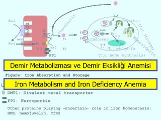
Cellular control of iron transport in duodenal enterocyte Brush border Basolateral DMT1 Ferroportin Fe(II) Fe(II) Fe(II) Fe(II) Fe(II) Hephaestin Fe reductase Fe(II) Fe(II) Fe(II) Labile iron Fe(II) Fe(II) pool Fe(III) Fe(III) Fe(II) Fe(II) Fe(II) Tf Fe(II) Fe(III) HCP1 Fe(III) Haem Endocytic Fe(III) Fe(III) Tf vesicle Fe(III) Fe(III) Fe(III) TfR ferritin Fe(III) Tf = transferrin; TfR = transferrin receptor.
Cellular control of iron transport in duodenal enterocyte Brush border Basolateral Hepcidin DMT1 Ferroportin Fe(II) Fe(II) Fe(II) Fe(II) Fe(II) Hephaestin Fe reductase Fe(II) Fe(II) Fe(II) Labile iron Fe(II) Fe(II) pool Fe(III) Fe(III) Fe(II) Fe(II) Fe(II) Tf Fe(II) Fe(III) HCP1 Fe(III) Haem Endocytic Fe(III) Fe(III) Tf vesicle Fe(III) Fe(III) Fe(III) TfR ferritin Fe(III) Tf = transferrin; TfR = transferrin receptor.
Stimulatory and inhibitory signals to hepcidin Hepcidin Erythropoiesis Low Fe stores Hypoxia Iron overload Inflammation L GDF15 TMPRSS6 HIF1-α H SMADs STAT-3 Ajioka RS, Prchal J. The Hematologist. 2008;5:(5)1.
Inherited IDA Andrews NC. Blood. 2008;112(2):219-30 .
Prevalence of iron deficiency anaemia (%) The population of Earth is estimated to be 6,993,000,000 ( US Census Bureau) – hence IDA may affect over 2 billion people worldwide DeMaeyer E, Adiels-Tegman M. World Health Stat Q. 1985;38:302-16.
Causes of iron deficiency Increased physiological requirements: growth, menses pregnancy EPO Limited supply: dietary malabsorption placental Blood loss menorrhagia GIT loss parasites other
Risk groups: 0 – 18yrs • Premature / sick infants • Certain ethnic groups • Aboriginals, immigrants • Growth phases • First 2 years • Adolescence • Excess loss • menses
Hypochromia Microcytosis Anisocytosis High RDW Typically low to normal RCC The Blood in IDA • Differential diagnosis: • Iron deficiency • Thalassaemia • Sideroblastic anaemias - rare • Lead poisoning - rare Hoffbrand AV, et al., editors. Essential haematology.5th ed. Malden, MA; Oxford: Blackwell, 2006.
Stages of iron deficiency Depleted iron stores Iron deficiency (normal Hb) Iron deficiency anaemia Serum ferritin Transferrin sat Erythrocyte ZPP Haemoglobin MCV % Hypo Serum TfR CHr CHr = haemoglobin content of reticulocytes; Hb = haemoglobin; Hypo = hypochromic erythrocytes; ZPP = zinc protoporphyrin. Modified after Brugnara C. Clin Chem. 2002;48:981-2.
Beware - iron studies • Serum iron is labile • high if haemochromatosis, enteric iron load, sideroblastic, aplastic, ineffective erythropoiesis • low if deficiency, infection, fasting, vit C def. • Transferrin affected by disease states • low in infection/inflammation, malignancy hypoproteinemic states, congenital def • high with OCP, pregnancy • TIBC calculated from Transferrin • Transferrin Saturation calculated from Se Fe and TIBC • Ferritin • high in acute phase; liver disease/injury, iron loading • low in deficiency, congenital (rare) • Interpretable in acute phase if know CRP • >100umol/L in CRF, chronic inflammation - Fe deficiency unlikely
Other indicators of iron status • Reticulocyte Hb ( CHr) • Indirect measure of iron available for new red cell production (few days) • Useful for diagnosis of deficiency and response to therapy (esp IV) • BUT not routinely available on all analysers or validated for all populations • Zinc Protoporphyrin • Old test and very sensitive to Fe deficiency • Accumulates in Fe deficiency and lead poisoning • Not readily available – referred test for many labs • Transferrin Receptor • Maintains cellular iron homeostasis • Increased production if iron deficient or if increased erythropoiesis • Useful marker of deficiency in states where there is confounding effect of inflammation / infection • Not helpful to discriminate thalassemia trait as levels overlap with those of iron deficiency • Available most labs
Case 1 – mohamid Hb 93, MCV 61 RDW 20 ( 11 – 15) CRP 24 Ferritin 32umol/l Iron deficient Tests depend on why ? diet ? bleeding ?? malabsorption Case 2 – mahali Hb 100, MCV 67 RDW 14.6 ( 11 – 15) CRP 28 Ferritin 159umol/l Thal trait iron deficiency unlikely Additional testing with haemoglobin studies Iron deficiency or thal trait? - A common conundrum
Iron deficiency vs thalassemia • Both reasonably common and can coexist • Assess for iron intake / malabsorption/ loss issues • Consider age • Iron issues peak in 0 – 4 and 10 – 16y • Ethnicity and family history may be helpful • ? RC indices • RDW, RCC, morphology ( stippling / targets++), dimorphism • Iron studies first line in most • Be aware of limitations and effects of acute phase • Interpretation with CRP helpful • Thalassemia testing if iron replete ( ? Post trial of iron) • Hb studies +/- family studies +/- alpha gene testing
Its iron deficiency! - BUT why is it present? Hb, MCV, Tf saturation, serum ferritin +/- sTfR Initial workup: Infants + “supply” cause Most others Category: Adolescents Detailed medical +/- gynaecological history +/- Occult blood Negative Positive Proceed to treatment GI workup GI = gastrointestinal.
GIT causes of IDA Coeliac disease IgA level and endomysial and TTG antibodies H. pylori infection Occult bleeding, competition for iron, interferes with acid production ( iron conversion) serology and urease breath test Worth thinking about especially in certain ethnic groups ( see next slide) Occult / overt bleeding Eg GOR / oesphagitis; Meckels; telengiectasia / angiodysplasia; portal HT; Inflammatory bowel disease Human Hb, calprotectin, endoscopy Iron transport defect Iron absorption challenge; genetic testing** Autoimmune atrophic gastritis Rare in children, association with H Pylori infection gastrin, parietal cell antibodies, anti-IF TTG = tissue transglutaminase;anti-IF = anti-intrinsic factor. .
Prevalence of H. pylori infection 100 80 60 Developing countries Developed countries 40 20 0 10 20 30 40 50 60 70 80 0 Prevalence (%) Age (years) Logan RP, Walker MM. BMJ. 2001;323:920-2.
Options for treatment of IDA Oral medications: tablets ferrous sulphate, gluconate or citrate containing ~ 50 mg elemental iron / tablet +/- vitamin C/ folate Oral medications: syrup ferrous sulphate – liquid 6mg elemental iron / ml Parenteral preparations - IV Venofer: iron saccharose 100 mg/5ml ampoule Maximum dose 1 ampoule Ferinject: iron carboxymaltose 100 mg/ 2ml or 500mg / 10ml vials ( dilute 100mg / 50ml N Saline) Various dosing protocols – Formula; <35kg -15mg/kg, >35kg – 500mg; 15mg/kg up to 1000mg maximum NOTE: Maximum weekly dose 1000mg Muñoz M, et al. J Clin Pathol. 2011;64:287-96.
Principles of IDA treatment Response rate to parenteral and oral iron is similar Difference between formulations mainly cost not quality Choice based on age / acceptance by patient Mostly trial oral replacement would precede IV iron Compliance important to consider Administration issues Consider degree of symptoms / tolerance to decide dose/ frequency and agent Duration of treatment should be very long At least 4 months for adequate repletion with standard oral dosing Response to iron is the ultimate test for IDA Hershko C, Skikne B. Semin Hematol. 2009;46:339-50.
Iron = 0mg / 5gm Iron = 0.17mg/5gm 10% absorption ~ 0.015mg/5gm ~1.5mg/90gm Iron = 6.5mg/5gm (spinach 0.9mg / cup fresh) if 5% absorption ~ 0.33mg / 5gm + Vitamin C ( and B12) Few studies (rats / humans) - improves iron status /non toxic - ? dose Food for thought?