Download
1 / 47
470 likes | 582 Views
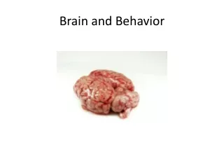
Brain and Behavior and Drugs: 1 st pt Chapter 3. In 1800, Franz Gall suggested, that bumps of the skull represented mental abilities. His theory though incorrect, nevertheless proposed different mental abilities were modular. History of the Mind. Phrenology. Bettman/ Corbis.

E N D
In 1800, Franz Gall suggested, that bumps of the skull represented mental abilities. His theory though incorrect, nevertheless proposed different mental abilities were modular. History of the Mind Phrenology Bettman/ Corbis
Neural Communication The body’s information system is built from billions of interconnected cells called neurons.
Neural Communication Neurobiologists and other investigators understand that information processing in humans and animals operate similarly. Note similarity of brain regions involved with information processing of similar kind.
Neuron A nerve cell or a neuron consists of many different parts.
Synapse Synapse [SIN-aps]ajunction between the axon tip of the sending neuron and the dendrite or cell body of the receiving neuron. This tiny gap is called the synaptic gap or cleft.
Neurotransmitters Neurotransmitters (chemicals) released from the sending neuron, travel across the synapse and bind to receptor sites on the receiving neuron, thereby influencing it to generate an action potential.
Lock & Key Mechanism Neurotransmitters bind to the receptors of the receiving neuron in a key-lock mechanism.
Nervous System Central Nervous System (CNS) Peripheral Nervous System (PNS)
The Nervous System Nervous System: Consists of all the nerve cells. It is the body’s speedy, electrochemical communication system. Central Nervous System (CNS): the brain and spinal cord. Peripheral Nervous System (PNS): the sensory and motor neurons that connect the central nervous system (CNS) to the rest of the body.
Kinds of Neurons Sensory Neurons carry incoming information from the sense receptors to the CNS. Motor Neuronscarry outgoing information from the CNS to muscles and glands. Interneurons connect the two neurons. Interneuron Neuron (Unipolar) Sensory Neuron (Bipolar) Motor Neuron (Multipolar)
Peripheral Nervous System Somatic Nervous System: The division of the peripheral nervous system that controls the body’s skeletal muscles. Autonomic Nervous System: Part of the PNS that controls the glands and other muscles.
The Nerves Nerves consist of neural “cables” containing many axons. They are part of the peripheral nervous system, and connect muscles, glands, and sense organs to the central nervous system.
Autonomic Nervous System (ANS) Sympathetic Nervous System: division of the ANS that arouses the body, mobilizing its energy in stressful situations. Parasympathetic Nervous System: division of the ANS that calms the body, conserving its energy.
Autonomic Nervous System (ANS) Sympathetic NS “Arouses” (fight-or-flight) Parasympathetic NS “Calms” (rest and digest)
Central Nervous System The Spinal Cord and Reflexes Simple Reflex
Central Nervous System The Brain and Neural Networks Interconnected neurons form networks in the brain. Theses networks are complex and modify with growth and experience. Complex Neural Network
Neural Communication Neurobiologists and other investigators understand that humans and animals operate similarly when processing information. Note the similarities in the above brain regions, which are all engaged in information processing.
Neuron The body’s information system is built from billions of interconnected cells called neurons which consist of many different parts.
Neurotransmitters Neurotransmitters (chemicals) released from the sending neuron travel across the synapse and bind to receptor sites on the receiving neuron, thereby influencing it to generate an action potential.
Reuptake Neurotransmitters in the synapse are reabsorbed into the sending neurons through the process of reuptake. This process applies the brakes on neurotransmitter action.
The Brain: Older Brain Structures The Brainstemisthe oldest part of the brain, beginning where the spinal cord swells and enters the skull. It is responsible for automatic survival functions.
The Brain: Older Brain Structures The Brainstemis responsible for automatic survival functions. The brain’s increasing complexity arises from new brain systems built on top of old. Within the brainstem are the oldest regions, the medulla and the reticular formation.
Brainstem The Medulla [muh-DUL-uh] is the base of the brainstem that controls heartbeat and breathing.
Brainstem The Thalamus [THAL-uh-muss] is the brain’s sensory switchboard, located on top of the brainstem. It directs messages to the sensory areas in the cortex and transmits replies to the cerebellum and medulla.
Brainstem Reticular Formationisa nerve network in the brainstem that plays an important role in controlling arousal.
Amygdala The Amygdala [ah-MIG-dah-la] is linked to the emotions of fear and anger.
Hypothalamus The Hypothalamus lies below (hypo) the thalamus. Directs several maintenance activities like eating, drinking, body temperature, and control of emotions.
Cerebellum The “little brain” attached to the rear of the brainstem. It helps coordinate voluntary movements and balance.
The Brain Techniques to Study the Brain A brain lesion experimentally destroys brain tissue to study animal behaviors after such destruction. Hubel (1990)
Clinical Observation Clinical observations have shed light on a number of brain disorders. Alterations in brain morphology due to neurological and psychiatric diseases are now being catalogued. Tom Landers/ Boston Globe
Electroencephalogram (EEG) An amplified recording of the electrical waves sweeping across the brain’s surface, measured by electrodes placed on the scalp. AJ Photo/ Photo Researchers, Inc.
PET Scan PET (positron emission tomography) Scan is a visual display of brain activity that detects a radioactive form of glucose while the brain performs a given task. Courtesy of National Brookhaven National Laboratories
MRI Scan MRI (magnetic resonance imaging) uses magnetic fields and radio waves to produce computer-generated images that distinguish among different types of brain tissue. Top images show ventricular enlargement in a schizophrenic patient. Bottom image shows brain regions when a participants lies. Both photos from Daniel Weinberger, M.D., CBDB, NIMH James Salzano/ Salzano Photo Lucy Reading/ Lucy Illustrations
The Limbic System The Limbic Systemisa doughnut-shaped system of neural structures at the border of the brainstem and cerebrum, associated with emotions such as fear, aggression and drives for food and sex. It includes the hippocampus, amygdala, and hypothalamus.
Reward Center Rats cross an electrified grid for self-stimulation when electrodes are placed in the reward (hypothalamus) center (top picture). When the limbic system is manipulated, a rat will navigate fields or climb up a tree (bottom picture). Sanjiv Talwar, SUNY Downstate
The Cerebral Cortex The intricate fabric of interconnected neural cells that covers the cerebral hemispheres. It is the body’s ultimate control and information processing center.
Structure of the Cortex Each brain hemisphere is divided into four lobes that are separated by prominent fissures. These lobes are the frontal lobe (forehead), parietal lobe (top to rear head), occipital lobe (back head) and temporal lobe (side of head).
Functions of the Cortex The Motor Cortex is the area at the rear of the frontal lobes that control voluntary movements. The Sensory Cortex (parietal cortex) receives information from skin surface and sense organs.
Motor & Sensory Cortex • Motor cortex controls voluntary muscle movements on the opposite side of the body. • Body parts requiring the most precise control occupy the greatest amount of cortical space. • Researchers have recorded messages from brain areas involved in planning and intention, leading to the testing of neural prosthetics for paralyzed patients. • Sensory cortex registers and processes body sensations. • The most sensitive body parts require the largest amount of space in the sensory cortex.
Visual Function Visual Function Auditory Function
Auditory Function The functional MRI scan shows the auditory cortex is active in patients who hallucinate.


![[PDF READ ONLINE] Psychopharmacology: Drugs, the Brain, and Behavior](https://cdn7.slideserve.com/12515541/slide1-dt.jpg)