Download

1 / 60
1.05k likes | 2.31k Views
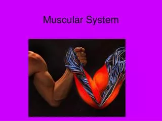
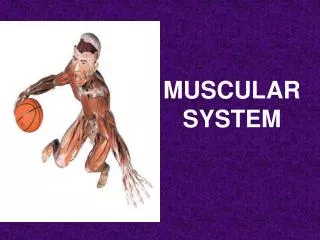
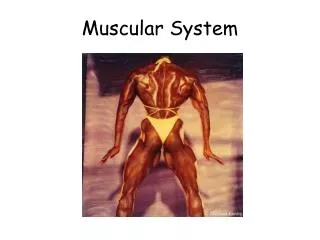
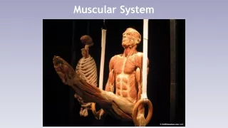
VERTEBRATE MUSCULAR SYSTEM. Mrs. Ofelia Solano Saludar Department of Natural Sciences University of St. La Salle Bacolod City. Muscle is a tissue; muscles are organs. HISTOLOGY : striated, cardiac, smooth FUNCTION : contraction

E N D
VERTEBRATE MUSCULAR SYSTEM Mrs. Ofelia Solano Saludar Department of Natural Sciences University of St. La Salle Bacolod City
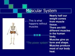
Muscle is a tissue; muscles are organs • HISTOLOGY: striated, cardiac, smooth • FUNCTION: contraction • locomotion: result of muscle action • posture determinant • orientation of body in the environment • heat production
EMBRYOGENESIS • Myotomesof epimere • Lateral mesoderm of hypomere • Somatic: body wall muscles • Splanchnic: smooth muscle of viscera
GROSS FEATURES OF SKELETAL MUSCLE Origin & insertion; Tendon; Aponeurosis; Fascia, Action
Various wrappings of connective tissues extend beyond the ends of the muscle fibers to connect with the periosteum of the bone: • Tendon- cordlike attachment consisting of extensions of a muscle's tough connective tissue sheath that anchor a muscle to its origin & insertion • Aponeurosis- thin flat sheet • Fascia- thin flat sheets of connective tissues that wrap and bind adjacent muscle groups • Raphe- junction of two muscles at a band of connective tissue to form a line of fusion, such as the linea alba
Agonists- primary action Antagonistic- oppose or resist the action of another muscle Synergistic- work together to produce a common effect
Names of skeletal muscles are based on: • action (e.g., levator scapulae) • direction of fibers (e.g., oblique) • location or position (e.g., superficial) • number of divisions (e.g., triceps) • shape (e.g., deltoid) • origin and/or insertion (e.g., iliocostalis) • size (e.g., major) • … or some combination of these
HISTOLOGY • STRIATED MUSCLE • Skeletal, voluntary muscles: axial, body wall & tail, hypobranchial & tongue, extrinsic eyeball, appendicular, branchiomeric or branchial muscles • Myofibrils are striated cylinders within syncytialmyofibers
MAJOR CATEGORIES OF STRIATED MUSCLES: somatic, visceral, branchiomeric somatic*
FIBER TYPE VARIATIONS WITHIN TWITCH FIBERS Muscles are mixtures of different fiber types; androgens & continued use result in increase in size & strength of muscle
SMOOTH MUSCLE TISSUE • Fusiform, uninucleate cells with myofibrils but without striations; occur in sheets • Two general types: • Unitary-has myogenic contraction to aid in sustaining the rhythmic movement of the organ • Multiunit- has neurogenic contraction, which requires action potentials sent by neurons
Lateral plate mesoderm in origin • Involuntary- innervated by ANS • Muscles of tubes, vessels, & hollow organs; intrinsic eyeball muscles; erectors of feathers & hair • Regulates internal body temperature
CARDIAC MUSCLE TISSUE • Heart muscle • Uninucleate, striated cells separated by intercalated disks • Lateral plate mesoderm in origin • Involuntary, self depolarizes (myogenic); ANS nerves modify its rhythmicity
AXIAL MUSCLES • Include the skeletal muscles of the trunk & tail • Are segmental because of their embryonic origin; arise from segmental mesodermalsomites • Metamerism is most evident in fishes and aquatic amphibians where the axial muscles are used in locomotion • Metamerism is obscured in tetrapods due to presence of paired appendages responsible for locomotion on land • Myotomesare separated by myosepta which serve as muscle origins & insertions • Myoseptum becomes indistinct in amniotes
Myotomes become divided by the horizontal skeletogenous septa into: • EPAXIALS-above the septum, dorsoflex spine • HYPAXIALS- below the septum, ventroflex spine • present in orbits as extrinsic eyeball muscles • extend forward beneath the pharynx as hypobranchial muscles & muscles of the tongue
JAWED FISHES EpaxialMuscles: • Innervated by dorsal rami of spinal nerves • Extend spine & some lateral bending • Extrinsic eye muscles (innervated by cranial nerves)
Hypaxial Muscles: • Innervated by ventral rami of spinal nerves • Ventroflexand lateral bending • Hypobranchialmuscles: hypaxialmuscles that migrated forward &come to lie on floor of pharynx, pectoral girdle to jaw; function in respiration & feeding, e.g. coracomandibularis
TETRAPODS Epaxialsare elongated bundles that extend through many body segments located below the expanded appendicular muscles; required to operate the limbs lie along vertebral column
Urodeles & some lizards - epaxials (DORSALIS TRUNCI) are still obviously metameric Anterior lateral musculature of a urodele (Ambystoma or tiger salamander)
Beginning with fishes, epaxial bundles split into longitudinal systems: long, short & segmented • Short & long bundles both arch & support the vertebral column • Extend from base of the skull to tip of the tail SHORT BUNDLES: • Extend from the 1st vertebrae to the skull (occipitals) • Short segmental muscles (intervertebrals) include several systems between various parts of the vertebrae & ribs, with each member extending only over one body segment • Connect processes of adjacent vertebrae • Tetrapodbundles perform same function as in fishes (side-to-side movements of vertebral column)
LONG BUNDLES: • Longissimusgroup- lies on transverse processes of vertebrae; includes the longest epaxial bundles: longissimusdorsi, longissimuscervicis, longissimuscapitis • Iliocostalisgroup- lateral to longissimus& spinalis; arises on ilium & inserts on dorsal ends of ribs or uncinate processes • Spinalisgroup- lies close to neural arches; connects spinousprocesses or transverse processes with those several vertebrae anteriorly
Hypaxials: • Muscles of lateral body wall: oblique (external & internal), transverse, & rectus muscles • Muscles that form longitudinal bands in roof of body cavity: subvertebral muscles • Hypaxialsof the abdomen have no myosepta & form broad sheets of muscle
OBLIQUE & TRANSVERSE MUSCLES Early amphibians & reptiles: ribs developed in myosepta along entire length of the trunk; Urodeles still have myosepta the length of the trunk, but ribs no longer form in all of them oes, obliquusexternussuperficialisoep, obliquusexternusprofundusoi, obliquusinternusta, transversusabdominis
Modern amniotes: myosepta & ribs are restricted to the thorax, hence abdominal muscles are not obviously segmented • Hypaxials are reduced in volume compared to fishes; support contents of abdomen & assist in respiration
RECTUS MUSCLES • Weakly developed in most fishes but stronger in tetrapods • Support ventral body wall, compresses abdomen, assist epaxialsin bending vertebral column • Consists of: rectus abdominis, cervicis, and geniohyoid in front of hyoid apparatus • Diaphragm – unique to mammals for breathing
INTERCOSTALSexternal & internal intercostal (respiration in amniotes) External intercostalmuscles Internal intercostalmuscles Ribs Intercartilaginous muscles Sternum Subcostal muscles Vertebral column
SUBVERTEBRAL MUSCLES • Underneath & against transverse processes of vertebrae • Includes the psoas & iliacus in the lumbar region & the longuscolliin the neck • Less developed in the thorax; none in the tail
ACTIONS OF SELECTED AXIAL MUSCLES Muscles of the back: Longissimusdorsi - extends vertebral column Iliocostalis - draws ribs together Multifidusspinae - extends vertebral column Spinalisdorsi - extends vertebral column Abdominal muscles: Rectus abdominis - compresses abdomen Internal oblique - compresses abdomen External oblique - constricts abdomen Internal oblique - constricts abdomen Respiratory muscles: Serratus - draw ribs cranially Scalenus - flexes the neck Diaphragm - separates the thoracic/abdominal cavities, functions in breathing Intercostals - protract/retract ribs
Arise from preoticsomitomeres • 6 voluntary muscles • Obliquesrotate eye along its transverse axis; rectus move eyes up, down, left, right; retractor in some • Retractor bulbipulls the eyeball further into the orbit to allow for coverage by the nictitating membrane (lacking in humans) • Innervated by the oculomotor nerve EYE MUSCLES
BRANCHIOMERIC MUSCLES • Develop from somitomeres & the myotomes caudal to those that produce the ocular muscles • Closely associated with the visceral skeleton so they are used in both breathing and feeding. • Perform the function of operating the jaw, opening and closing the spiracle • Primitively had a levator & a constrictor muscle series; in present vertebrates coracobranchials, subarcualsandventral transversals are added • May be subdivided based on what visceral arch they are associated with
Muscles of the Mandibular Arch: • FISHES- operate the jaws: adductor mandibulae&intermandibularis (mylohyoidin mammals) • TETRAPODS- muscles of 1st arch still operate jaws; adductors of mandible: masseter& temporalis, pterygoid, anterior belly of digastric, tensor palati&tensor tympani of mammals Muscles of the Hyoid Arch: • Constrictors:interhyoideus (posterior belly of digastric) & constrictor colliof reptiles & birds (platysma & facial muscles in mammals) • Levatorsbecome depressor mandibulae& stapedius • In fishes, muscles become reduced because the operculum plays important role in respiration
Muscles of 3rd & successive arches: • Levators: cucullarisof gill-bearing vertebrates become the trapezius & sternocleidomastoid of tetrapods • Constrictors have no representatives in tetrapods • In tetrapods, primary muscles include:stylopharyngeus (Arch III) - used for swallowing • Remaining arches give rise to intrinsic muscles of the larynx or ‘voicebox’
TONGUE MUSCLES • Hypobranchial- ventral muscles of the head and trunk region that perform functions associated with jaw and tongue movement • Extend forward from pectoral girdle & insert on mandible, hyoid, & gill cartilages • Strengthen floor of pharynx • Assist branchiomeric muscles in elevating floor of mouth, lowering jaw, & extending gill pouches
Fishes- associated with feeding and breathing: • Coracoarcuals- opens mouth • Coracomandibular- opens mouth • Coracohyoid- helps in feeding • Coracobranchial- helps in swallowing • Tetrapods- associated with the hyoid apparatus & tongue • Tongue muscles: hyoglossus, styloglossus, genioglossus (also speech & sound production) • Geniohyoid: draws hyoid cranially • Sternohyoid: draws hyoid posteriorly • Sternothyroid: draws larynx caudally • Tongue of amniotes is a 'sac' anchored to hyoid skeleton & filled with hypobranchial muscle
Hypobranchialsending in "hyoid" stabilize hyoid and larynx; e.g. geniohyoid, sternohyoid, sternothyroid, thyrohyoid • Those beginning or ending with "thyro" are attached to the larynx; e.g. thyrohyoid • Those ending with “glossus” or start with “lingu” are tongue muscles, e.g. lingualis, styloglossus
APPENDICULAR MUSCLES • Muscles of girdles and appendages • Move fins or limbs • Innervated by ventral ramus of spinal nerves • Two types based on origin: • Extrinsic- originate on axial skeleton or fascia or trunk & insert on girdles or limbs • Intrinsic- originate on girdle or proximal skeletal elements of appendage & insert on more distal elements
FISHES • Appendicularmuscles serve mostly as stabilizers • Intrinsic muscles are limited in number and undifferentiated • Originated as extensions of hypaxials of body wall • Paired fins are appendicular (from myotome)
Median dorsal & ventral fins are NOT appendicular, from myotomeof epaxials & hypaxialsrespectively • Dorsal mass on paired fins are extensors or abductors • Ventral mass on paired fins are flexors or adductors
TETRAPODS • Appendicularmuscles are much more complicated than in fish • Greater leverage required for locomotion on land • Jointed appendages (as opposed to fins) require complex muscles
INTRINSIC (PRIMARY) APPENDICULAR MUSCLES • Form from blastemas within the limb bud • Amphibians- much more complex than in fish • Reptiles- more numerous & diverse than in amphibians; better support of body & increased mobility of distal segments of the limbs • Mammals - similar to reptiles but more diverse
BIRDS • Intrinsic musculature is reduced • Pectoralis(humerus adductor), is the largest flight muscle that lowers wing • Supracoracoideuselevates wing
EXTRINSIC APPENDICULAR MUSCLES • Dorsal groupof the forelimbs (e.g., trapezius and latissimusdorsi) arise on: • fascia of trunk in lower tetrapods • skull, vertebral column, & ribs to a point well behind the scapula in higher tetrapods & converge on the girdle & limb • Ventral group (e.g., pectoralis) arises on sternum & coracoid, & converge on limb • RESULT = pectoral girdle & limb are joined to trunk by extrinsic appendicular muscles