Download
1 / 18
240 likes | 1.57k Views
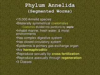
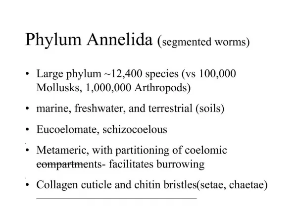
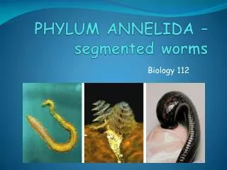
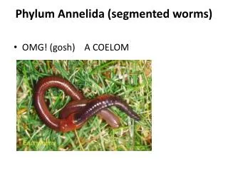
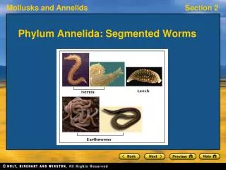
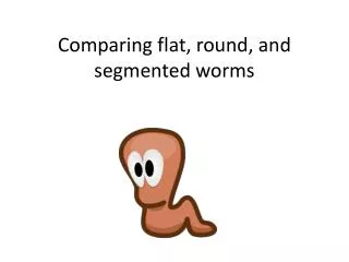
Comparing flat, round, and segmented worms. Platyhelmenthes cross section (flat worm). Platyhelmenthes. Nematoda cross section (round worm) . Nematoda. Annelida cross section (segmented worm). Annelida. Tissues.

E N D
Tissues • Animal body plans also vary according to the organization of the animal’s tissues • Tissues are collections of specialized cells isolated from other tissues by membranous layers • During development, three germ layers give rise to the tissues and organs of the animal embryo
Ectoderm is the germ layer covering the embryo’s surface • Endoderm is the innermost germ layer and lines the developing digestive tube, called the archenteron • Diploblastic animals have ectoderm and endoderm • Triploblastic animals also have an intervening mesoderm layer; these include all bilaterians
Body Cavities • Most triploblastic animals possess a body cavity • A true body cavity is called a coelom and is derived from mesoderm • Coelomates are animals that possess a true coelom
Fig. 32-8 Coelom Body covering (from ectoderm) Tissue layer lining coelom and suspending internal organs (from mesoderm) Digestive tract (from endoderm) (a) Coelomate Body covering (from ectoderm) Pseudocoelom Muscle layer (from mesoderm) Digestive tract (from endoderm) (b) Pseudocoelomate Body covering (from ectoderm) Tissue- filled region (from mesoderm) Wall of digestive cavity (from endoderm) (c) Acoelomate
Fig. 32-8 Coelom Body covering (from ectoderm) Tissue layer lining coelom and suspending internal organs (from mesoderm) Digestive tract (from endoderm) (a) Coelomate Body covering (from ectoderm) Pseudocoelom Muscle layer (from mesoderm) Digestive tract (from endoderm) (b) Pseudocoelomate Body covering (from ectoderm) Tissue- filled region (from mesoderm) Wall of digestive cavity (from endoderm) (c) Acoelomate
Fig. 32-8a Coelom Body covering (from ectoderm) Tissue layer lining coelom and suspending internal organs (from mesoderm) Digestive tract (from endoderm) (a) Coelomate
A pseudocoelom is a body cavity derived from the mesoderm and endoderm • Triploblastic animals that possess a pseudocoelom are called pseudocoelomates
Fig. 32-8b Body covering (from ectoderm) Pseudocoelom Muscle layer (from mesoderm) Digestive tract (from endoderm) (b) Pseudocoelomate
Triploblastic animals that lack a body cavity are called acoelomates
Fig. 32-8c Body covering (from ectoderm) Tissue- filled region (from mesoderm) Wall of digestive cavity (from endoderm) (c) Acoelomate
Microscopic observation • Use the prepared slides of the three different worm types. • Divide a sheet of printer paper into three columns, and label each with the name of a different worm. • Draw each from the slide you see, and label as many parts as you can identify. Don’t guess. • Are you able to identify the gender? • Label the three different tissue layers. • Label the body cavity (if present).