Download
1 / 39
390 likes | 586 Views
A thirty three years-old man presented with dyspnea, fever and cough. Dr. Ahmet Bircan University of Suleyman Demirel Faculty of Medicine Dept. of Pulmonary Medicine, Isparta. Turkish Thoracic Society 13 th Annual Congress, 2010, Istanbul. Case presentation. 33 years-old, man Symptoms:

E N D
A thirty three years-old man presented with dyspnea, fever and cough Dr. Ahmet Bircan University of Suleyman Demirel Faculty of Medicine Dept. of Pulmonary Medicine, Isparta Turkish Thoracic Society 13th Annual Congress, 2010, Istanbul
Case presentation • 33 years-old, man • Symptoms: • Dyspnea, fever, cough and weight loss • History: • No complaints until 45 days before his admission • Symptoms; had started 45 days ago • Cough; nonproductive • Fever; once every 2-3 nights, without taking his temperature • Weight loss; inappetence, malaise and weight loss (3 kg) (+) in the last 2 months
No co-morbidity • Smoking: never • Family story: nothing special
Physical Exam • Good condition • Blood pressure: 100/60 mmHg, • Pulse: 94/min., • Temperature: 36 0C, • Respiratory rate: 24/min., • Respiratory system exam: • In oscultation, fine crackles were found bilaterallyat the base of hemithorax • All the other systems were normal
Hemogram Leukocyte: 12.000/mm3, Neutrophile, 85.6%, Lymphocyte, 8.5% Hemoglobin: 17.2 g/dl, Hematocrit: %47 Platelet: 307.000/mm3, ESR: 3 mm/h CRP: 34.8 mg/L Biochemistry AST: 45 U/L (N:10-40) LDH: 517 U/L, (N:150-290) All other biochemical analyses were in normal limits Laboratory findings-1
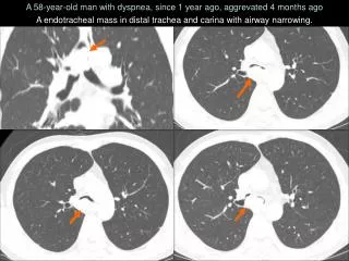
Thorax HRCT diffuse, multiple, small andill-defined centrilobular-acinar nodules and patchy ground-glass opacities in both lungs
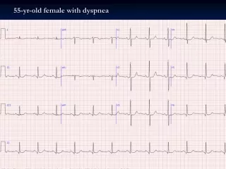
ABG analysis, breathing in room air Pulmonary function tests Laboratory findings-2
Laboratory findings-3 • AFB smear (-) • From inducted sputum and BAL • ANA: (-) • RF: N • Compleman: N • ACE: N • IgG: 2310 • IgA, IgE ve IgM; N
What is your diagnosis with these findings? • Atypical pneumonia • Miliary tuberculosis • Sarcoidosis • Hypersensitivity pneumonitis • Silicosis • Organic toxic dust sydrome • Silo filler’s disease • Reactive airway dysfunction syndrome (RADS)
What is your next step in diagnostic approach? • More information about history • Serological tests for M.pneumoniae • Galium scintigraphy • Bronchoscopy (BAL,TBB) • Reversibility test • Surgical biopsy
No tb contacts in his family • Occupation: • 10 years ago, he had worked in a textile factory for 2 years, with no respiratory symptoms • working as a security staff in a bank for 2 years • Hobby: • A pigeon breeder for 7 years
What is the next procedure? • FOB+BAL • FOB+TBNA • FOB+BAL+TBB • EBUS • Provocation tests • VATS biopsy • Open lung biopsy
Bronchoscopic examination revealed no endobronchial patology • BAL was done • TBBs were obtained from LLL • Due to technical errors cytologic analyses of the BAL fluid did not performed
Hypersensitivity pneumonitis extrinsic allergic alveolitis Pigeon breeders disease Pigeon, turkey, duck, parrots, cannaries, chickens, geese
History of symptoms compatible with HP Confirmation of exposure to the offending agent by history, serum and/or BAL fluid antibody. Characteristic radiological findings BAL fluid lymphocytosis Compatible histological changes Positive natural challenge or by controlled inhalational challenge. Basilar crackles. Decreased diffusion capacity. Arterial hypoxaemia Diagnostic criteria Major (at least 4) Minor (at least 2) Schuyler M. The diagnosis of hypersensitivity pneumonitis. Chest 1997; 111: 534–536.
Predictors of Hypersensitivity pnömonitis • Presence of all 6 probability of HP was 98% Lacasse Y, et al. AJRCCM 2003; 168: 952-8.
Aetiology • More than 200 types of HP were described • Clinical, radiological and pathological similarities of these types of HP suggest us an common pathogenesis. • Thermophilic actinomyches..Farmers Lung • Penicillium spp… Humidifier Lung • Animal proteins….Pigeon Breeders Lung • Animal proteins…Bird Fancier’s Lung • Fungus…Malt Workers Lung • MAC…..Hot Tub Lung • And many many more……
What is most common form of HP? • Farmer lung disease • Pigeon breeder disease • Bagassosis • Japanese summer type HP • Drug induced HP • HP due to chemical compounds
Epidemiology • Prevalance of HP is variable in different population • Geographical features, climate and production of industrial crops • In Turkey, FLD is more frequently seen in the region of East Anatolia (livestock) and Black Sea (production of nuts) • The most frequent types of HP in European countries are HPs seen in pigeon breeders and in farmers • Among farmers----- FLD 1-19%; • Among pigeon breeders------ PBD 6-20% • 80-95% of patients are nonsmoker. Prognosis is poor in smokers
Histopathology Acute HP • Inflammatory interstitialinfiltrate • Scattered poorly formed non-caseating granulomas and multinucleated giant cells • Cellular Bronchiolitis • These features seen in up to 75% of cases • Vasculitis and eosinophils are not present • Subacute HP • Interstitiel mononuclear infiltrate • chronic HP • Interstitiel fibrosis Takemura T, et al. Curr Opinion Pulm Med 2008,14:440–54.
Pathogenesis • Type III humoral mechanism • IgG (IgA or IgM) • Complex with inhaled antigen to fix complement • Stimulate alveolar macrophages to secrete inflammatory mediators • Neutrophilic chemotactic factors • Proteases • Reactive oxygen intermediates • IL-8
Pathogenesis • Type IV cell mediated response • Occurs with ongoing exposure to Ag • Activated macrophages secrete IL-12 • Promotes CD4+ Th0 lymphocytes to Th1 • IL-1 and TNF-alpha stimulate Th1 cells to produce IFN-gamma(a key mediator) • IL-10 (counter-regulatory mediator) • Other Chemokines • IL-8 and MCP-1 • Produces by alveolar macrophages • Chemoattractant to CD8+ lymphocytes into the lung • MIP-1 • Produced by CD8+ lymphocytes & activated macrophages • Facilitate the differentiation of alveolar macrophages into epithelioid cells & multinucleated giant cells
Pathogenesis • Progression to fibrosis • TGF-Beta • Fibroblast chemoattractant, collagen production • TNF-alpha • Stimulate the proliferation of collagen producing fibroblasts in the interstitial space through TGF-B mediated pathways
Clinical presentation • Affected by multiple factors • Antigenic feature of organic particle, it’s size and concentration • Frequency and intensity of antigen exposure • Host immune response • Co-infections • Classically, consisted of acute, subacute and chronic forms. • Alternate classification schemes have been proposedbecause of: • clinical course is so highly variable • acute forms of HSP do not necessarily evolve into a chronic form of the disease
Classification Hypersensitivity Pneumonitis: A Hypothesis Data obtained from a large prospective multicenter cohort study (the HP Study)168 patients Aktive (n=41) • Recurrent symptoms • Normal CXR Sequela (n=127) • Clubbing • Hypoxemia • Restrictive PFT • HRCT reveals fibrosis P<0.0001 • In each group, frequency of nodular opacities were similar • Subacute HP is difficult to define Lacasse Y, et al. Classification of hypersensitivity pneumonitis: a hypothesis. Int Arch Allergy Immunol 2009:149:161-6.
Acute form Subacute form Chronic form Dyspnea, fever, cough and weight loss for a duration of six weeks Bibasillar crackles Centrilobularnodulles Which clinical form is consistent with our case?
Acute form • Occur in previously sensitized patients with intermittent high intensity antigenic exposure • Influenza like symptoms; dyspnea, nonproductive cough, fever, chills, myalgia and headache <1 month • Begin 2-9 h after exposure • Peak during 6-24 h • Usually resolves 24-48 h • Bibasilar crakles(+) 47y, M, bird-related HP, bilaterally asymetric ground glass opacity Silva, C. I. S. et al. Am. J. Roentgenol. 2007;188:334-344 38 y, W, antigen? Matar LD, AJR. 2000; 177: 1601-6.
Subacute form • Small amounts of antigen exposure for a long period • More insidious onset (severalweeks- month) • Is marked by cough and dyspnea, leading to hospitalization • P.E. reveals bibasilar crackles (+) and dyspnea on exertion (+) • Removal of the patient from the offending environment improves the symptoms Silva, C. I. S. et al. Am. J. Roentgenol. 2007;188:334-344
Chronic form • Occurs in 5% of HP patients • Slowly progressive dyspnea on exertion • Cough, malaise, weight loss • Removal of the patient from the offending environment does not improve the symptoms • Precipitating Ab may or may not be present • Interstitial fibrosis • Clubbing (+), in 20-50 %, poor prognosis • PHT, Cor pulmonale
Chronic form 56y, M, isocyanate compound, traction bronchiectasis, 77y, M, PBD, reticulonodular inf. Silva CIS, et al. Am J Roentgenol. 2007;188:334-44. 44y, M, FLD, emphysema
BAL • 3-5 fold increase in cell count • Neutrophils may predominate in lavage fluid if performed within 48 hours of acute exposure • Lymphocytic alveolitis (CD8+ T-lymphocytes ),>5 days • CD4+/CD8+< 1 • Type of HP • Duration of antigen exposure • Time of taking BAL • Cilinical presantation • in chronic form CD4+ • in acute form CD8+ • Smoking • smokers CD4+ • Increase IgG, IgM, IgA
Lung biopsy • TBB is of limited usefulness for the diagnosis • Surgical Lung biopsy • Diagnostic yield 34-100 % • Treatment alteration 46-75 % • Selection of patients • Timing of the procedure • Expertise of the attending pathologist • It should be reserved for rare cases with puzzling clinical presentation and when the clinical course or response to therapy is unusual Lung Biopsy • (This recommendation is not based on evidence) Girard M, Lacasse Y and Cormier Y. Allergy 2009: 64: 322–334
What is the most appropriate treatment option for this patient? • Removal of patients from antigenic exposure is sufficient • Removal of patients from antigenic exposure + inhaled CS • Removal of patients from antigenic exposure + oral CS • Removal of patients from antigenic exposure + oral CS + immunosupressive treatment
Treatment-preventing The most important steps • Making early diagnosis • Avoiding recurrent exposures • Decreasing the incidence of occupational risks; • Improved fresh air ventilation • Medical surveillance/restriction • Adapting modern agricultural practices • Cleaning habitat in home-related HP
Treatment • Removal of patients from antigenic exposure is generally sufficient • Oral corticosteroids; • Provides symptom control in acute/subacute forms • Does not influence long-term prognosis • 0.5-1 mg/kg/day prednisolone, until objective improvement occurs • 10-15 mg/day maintenance, for 6 months • Improvement has been reported with inhaled steroids in subacute HP, but studies are scanty
Take home messages • HP is an immunologically mediated lung disease mediated primarily by T-cell responses to inhaled antigens. • The diagnosis requires careful history, appropriate laboratory tests, and lung biopsy in selected patients. • Avoidance of exposure is associated with a good prognosis and corticosteroids are indicated in severely symptomatic patients • Because of constantly changing environmental exposures, new examples of HP are being described, and represent an ongoing challenge in patients withILD.