Download
1 / 31
530 likes | 2.02k Views
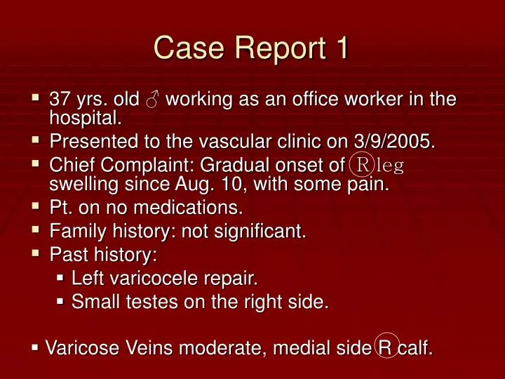
Varicose Veins moderate, medial side R calf. Case Report 1. 37 yrs. old ♂ working as an office worker in the hospital. Presented to the vascular clinic on 3/9/2005. Chief Complaint: Gradual onset of R leg swelling since Aug. 10, with some pain. Pt. on no medications.

E N D
Varicose Veins moderate, medial side R calf. Case Report 1 • 37 yrs. old ♂ working as an office worker in the hospital. • Presented to the vascular clinic on 3/9/2005. • Chief Complaint: Gradual onset of R leg swelling since Aug. 10, with some pain. • Pt. on no medications. • Family history: not significant. • Past history: • Left varicocele repair. • Small testes on the right side.
Case Report 2 • Telangiectasia (Chronic dilation of groups of capillaries causing elevated dark red blotches on the skin). • No Skin Changes or ulcerations • Doppler Report: • Distention of branches of the long saphenous veins (pretibial). • Clinically nothing is wrong with the short saphenous vein.
Anatomy and Physiology 1 • Blood flow from the legs • Blood is pumped from the heart to the legs through arteries. Once it has given up the oxygen and nutrients it was carrying, blood returns towards the heart through the veins. To do this from the legs, blood in the veins must flow upwards, against gravity. One-way valves inside the veins prevent the blood from flowing backwards. • The muscles in the legs help this flow. Each time the calf and thigh muscles contract when walking, veins deep inside the leg are squeezed. The valves ensure the blood travels upwards. • Surface and deep veins • Blood from the outer layers of the leg collects into veins near to the surface. These surface, or superficial, veins are connected to the deeper veins inside the leg by "perforator" veins. When blood does not flow properly from the surface veins to the deep veins, pressure can build up in the surface veins. This results in blood pooling and the visible sign of varicose veins.
Anatomy and Physiology 2 • Many veins, particularly those in the arms and legs, have one-way valves. Each valve consists of two flaps (cusps or leaflets) with edges that meet. Blood, as it moves toward the heart, pushes the cusps open like a pair of one-way swinging doors. • If gravity or muscle contractions try to pull the blood backward or if blood begins to back up in a vein, the cusps are pushed closed, preventing backward flow. Thus, valves help return blood to the heart—by opening when the blood flows toward the heart and closing when it tries to flow backward.
Anatomy and Physiology 3 • The legs contain superficial veins, located in the fatty layer under the skin, and deep veins, located in the muscles. Short veins, called connecting veins, link the superficial and deep veins.
Anatomy and Physiology 4 • The deep veins play a major role in propelling blood upward. The one-way valves in deep veins prevent blood from flowing backward, and the muscles surrounding the deep veins compress them, helping force the blood upward. • The powerful calf muscles are particularly important, forcefully compressing the deep veins with every step. The deep veins carry 90% or more of the blood from the legs toward the heart.
Anatomy and Physiology 5 • The One-way valves consist of two flaps (cusps or leaflets) with edges that meet. These valves help veins return blood to the heart. Blood, as it moves toward the heart, pushes the cusps open like a pair of oneway swinging doors (shown on the left). If gravity momentarily pulls the blood backward or if blood begins to back up in a vein, the cusps are immediately pushed closed, preventing backward flow.
Anatomy and Physiology 6 • Superficial veins play only a minor role in carrying blood to the heart. They have the same type of valves as deep veins, but they are not surrounded by muscle. • Thus, blood in the superficial veins is not forced upward by the squeezing action of muscles, and it flows more slowly than blood in the deep veins. Much of the blood that flows up the superficial veins is diverted into the deep veins through the many connecting veins between the deep and superficial veins. • Valves in the connecting veins allow blood to flow from the superficial veins into the deep veins but not vice versa.
Pathology 1 • Varicose Veins occurs as a result of: Abnormal communication between the deep and superficial venous systems. • The process usually begins with failure of the valve at the sapheno-femoral junction. • An uninterrupted column of the blood from the heart progressively dilates the vein down the leg.
Pathology 2 • Women are affected about six times more often than men, with the majority of varicose veins developing during or soon after the second or third pregnancy. • ↑ Progesterone → : • changes in the structure of collagen ( which may never recover!). • Smooth muscle relaxation. • ↑ Pressure on the pelvic veins by the enlarging uterus → Restriction of the venous return.
Pathology 3 • Hereditary factors appear to play a part, especially in men, particularly those who develop varicose veins in their teens.Predisposing anatomical factors may include: • Congenital lack of valves in the iliac veins. • Abnormal vein wall elasticity. • Rarely: • Multiple congenital arteriovenous fistulae (Klippel-Trynlawnay Syndrome “giganism of the lower limb and often venous ulceration”).
Signs and Symptoms + Common Complains • Aching legs, usually after standing all day. • Poor cosmetic appearance. • Varicose eczema or ulcers / Itching of the skin of the legs. • Recurrent superficial thrombophlebitis. • Ankle edema. • Fear of future leg ulcers “like my mother had!” • Worry about varicosities bleeding particularly if traumatized.
Thrombophlebitis: • Inflammation of the wall of the vein with secondary thrombosis occuring within the affected segment of the vein. It may involve superficial or deep veins of the legs. • DVT →PE
Signs and Symptoms + Common Complains 2 • Patients may present with acute varicose complications, including variceal bleeding, new onset of dermatitis, thrombophlebitis, cellulitis, and ulceration. • Patients may also consult a physician because of worsening chronic symptoms or for a variety of other reasons. • Some are seeking advice on the medical implications of varicose veins.
Venous History • The venous history should also include the following elements: • History of venous insufficiency (eg, date of onset of visible abnormal vessels, date of onset of any symptoms, any known prior venous diagnoses, any history of pregnancy-related varices) • Presence or absence of predisposing factors (eg, heredity, trauma to the legs, occupational prolonged standing, sports participation) • History of edema (eg, date of onset, predisposing factors, site, intensity, hardness, modification after a night's rest) • History of any prior evaluation of or treatment for venous disease (eg, medications, injections, surgery, compression) • History of superficial or deep thrombophlebitis (eg, date of onset, site, predisposing factors, sequelae) • History of any other vascular disease (eg, peripheral arterial disease, coronary artery disease, lymphedema, lymphangitis) • Family history of vascular disease of any type
Also in history taking… • Ask carefully about symptoms that would indicate a cause of the varicose veins, especially pregnancy and abdominal symptoms from pelvic tumors →Systems Review? • Previous History: • Duration of the V.V.? • Have had any form of treatment for the V.V. before?
Epidemiology • Age, V.V. affects all age groups but: • Young and middle aged women are the most common sufferers. • In children: congenital vascular abnormality. • Sex: Women are affected 6 times more than men. • Ethnic Group: V.Vs. were used to be less common in Africa and the Far East. Now? Why? • Occupation: jobs that involve standing up for long periods. So :don’t forget to ask about when taking history. But : causes ↔ exacerbates ?
Physical Examination 1 • Inspection: • Look for large visible veins. Record their site, extent and size. • Look at the skin for signs of chronic venous hypertension, eczema and ulceration.
Physical Examination 2 • Palpation: • Texture of the skin and subcutaneous tissue of the lower part of the leg. • Pitting edema? Thickening? Redness? Tenderness? → Chronic Venous Hypertension (lipodermatosclerosis: caused by a progressive sclerosis of the skin and subcutaneius fat by fibrin diposition, tissue death and scarring). • Feel along the course of the veins, particularly behind the medial border of the tibia→ Tender defects in the deep fascia? • Feel for pulse impulse in the groin? • Trenlenburg’s test.
Trendlenburg’s Test • Careful physical examination permits the identification of the level of reflux in most patients with peripheral varicosities. • The leg is raised to drain the venous system, and a venous compression tourniquet is placed at the level to be tested. When the leg is lowered, the deep veins fill quickly. • If there are incompetent perforators below the level of the tourniquet, the superficial varicosities below that level will also fill quickly. • If there is reflux from above the level being tested, release of the tourniquet will cause immediate visible distention of the superficial varicosities below.
Physical Examination 3 • Percussion : check for conduction of percussion impulse up and down the vein. • Auscultation ? • General Examination: • Abdomin • PR • Vaginal Examination • Examination of the testes • Harvey’s test (test the direction of the flow in a vein by placing two fingers on the vein, sliding one finger along the vein to empty it and then releasing one finger and inspection to see which way the empty segment fills).
Imaging Investigations 1 • Indications: • Recurrent V.V. • Short Saphenous V.V. • Where there is suspicion of deep venous pathology, for example previous DVT or skin changes of chronic venous insufficiency. • Atypical Distribution.
Investigations 2 • Color-flow Duplex Ultrasound Imaging • Color-flow imaging has become the standard modality for the evaluation of patients with venous disease of any type. The addition of color-coded flow information overlying a standard gray-scale ultrasound image allows rapid and accurate mapping of both deep and superficial veins, with identification of any flow disturbance (such as DVT) and of all areas of valvular incompetence and venous reflux. Arterial evaluation is also facilitated by color-flow imaging, especially with newer machines which offer 'Triplex' imaging with simultaneous gray-scale, color-flow, and M-mode doppler sampling information all on the same screen.
Investigations 3 • Contrast Venography • Radiopaque contrast material is injected into a distal foot vein, and tourniquets are placed so as to occlude the superficial venous system and force contrast into the deep venous system, permitting x-ray evaluation of the entire deep venous system. • Contrast venography was once the mainstay of the diagnostic workup for deep venous disease, but is now being replaced by duplex ultrasound imaging, which is of equal or greater sensitivity in the symptomatic patient. • Contrast venography continues to offer a higher sensitivity when used as a screening exam in a high-risk population, such as in patients with pulmonary embolism but with no leg symptoms.
Treatment 1 • Indications for surgery: • Cosmetic. • To prevent future complications. • Aching legs after standing relieved by elevation or in bed at night, particularly with unilateral ankle edema. • hemorrhage from a varicose vein. • Superficial thrombophlebitis. • Varicose skin changes. • All of these can be treated with support bandages or stockings, but surgery is often preferable.
Treatment 2 • Injection Sclerotherapy • The injection of a mild sclerosing agent is the initial treatment method of choice for most patients with peripheral venous varicosities and telangiectasias. Local tissue damage and discomfort are minimal because a #30 or #33 gauge needle is used. It may be used for varicosities and telangiectasias of any size, and is well-tolerated even where tiny facial vessels must be treated. A large varicosity that has failed treatment or has recurred after a course of injection/compression sclerotherapy may respond to a second course of sclerotherapy or it may require surgical ligation or even stripping.
Treatment 3 • High Saphenous Ligation • Ligation of an incompetent saphenofemoral junction may be performed in the office under local anaesthesia, and is often used in combination with compression sclerotherapy to treat a large varicosity with significant retrograde flow originating at the level of the saphenofemoral junction. Ligation of the site of inflow greatly increases the success of injection sclerotherapy along the entire length of the vessel. Ligation without sclerotherapy is rarely effective, because most patients have, or will develop, additional incompetent perforating vessels below the level of the saphenofemoral junction.
Treatment 4 • Surgical Stripping • Vein stripping, or surgically pulling the vessels from the body through a series of incisions, is the traditional surgical means of managing significant varicosities. It is the most invasive approach, has the most complications, requires the longest recuperation time, and leaves the most scarring. It should be performed in a surgical suite, and sometimes requires general anaesthesia. It has the lowest recurrence rate of all treatment modalities, but recurrences may occur even when the procedure is performed perfectly. Stripping does not improve many telangiectasias even when they communicate with a stripped varicosity.
Case Management • Q: How was the case which was mentioned in the beginning of this presentation managed ?? • A: By: Surgical Stripping.