Download
1 / 29
310 likes | 618 Views
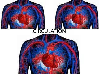
Circulation. Capillary. Red blood cell. Circulatory System. Necessary for large animals Can’t use diffusion System must have close connection with tissues Capillaries are microscopic blood vessels They form an intricate network among the tissue cells

E N D
Capillary Redbloodcell Circulatory System • Necessary for large animals • Can’t use diffusion • System must have close connection with tissues • Capillaries are microscopic blood vessels • They form an intricate network among the tissue cells • No substance has to diffuse far to enter or leave a cell
Capillary Diffusion ofmolecules INTERSTITIALFLUID Tissuecell Circulatory System
Circulatory Systems • Most animals have a separate circulatory system • Open circulatory system- • Invertebrates • A heart pumps blood through open-ended vessels into spaces between cells • Diffuse directly from blood into body cells • No distinction between blood and interstitial fluid
Tubularheart Pores Open Circulatory System
Closed Circulatory System • Blood is confined to the vessels so there is a difference between it and interstitial fluid • Most vertebrates have this cardiovascular system • 3 kinds of vessels • Arteries- away from heart • Veins- return blood to heart • Capillaries- between arteries and veins in organs
Capillary beds Arteriole Artery(O2-rich blood) Venule Vein Atrium Heart Artery(O2-poor blood) Ventricle Gillcapillaries Closed Circulatory System
The Human Heart • About the size of a clenched fist • Made of mostly cardiac muscle tissue • Atria- Collect blood returning to the heart and transport to ventricles • Ventricles- pump blood to all other bodily organs • Walls of left ventricle stronger for increased pump pressure • Valves regulate direction of blood flow
Pulmonaryartery Aorta Pulmonaryartery Superiorvena cava LEFTATRIUM RIGHTATRIUM Pulmonaryveins Pulmonaryveins Semilunarvalve Semilunarvalve Atrioventricularvalve Atrioventricularvalve Inferiorvena cava RIGHTVENTRICLE LEFTVENTRICLE The Human Heart
Circulation • Right ventricle (1) pumps blood to lungs through two pulmonary arteries (2). • Blood flows through capillaries (3) in the lungs, lets off CO2 and gets O2 • Blood flows back to the left atrium (4) through pulmonary artery • Blood flows from left atrium to left ventricle (5) • Blood leaves ventricle through aorta (6)
Circulation • Large arteries branch from the and lead to head and arms (7)and to the abdominal cavity and legs (8) • Oxygen-poor blood is pumped back into the superior vena cava(9)from the arms and the inferior vena cava (10) from the legs • The two large veins dump their blood back into the right atrium (11) • Process starts all over again
Superiorvena cava 7 Capillaries of Head and arms Pulmonaryartery Pulmonaryartery Capillariesof right lung Capillariesof left lung Aorta 9 6 2 3 3 4 11 Pulmonaryvein Pulmonaryvein 5 LEFT ATRIUM 1 RIGHT ATRIUM LEFT VENTRICLE RIGHT VENTRICLE 10 Aorta Inferiorvena cava Capillaries ofabdominal organsand legs 8
Structure of Blood Vessels • Capillaries- • Very thin walls, a single layer of epithelial cells • Very smooth • Arteries and veins • Very think walls • have smooth muscle and connective tissue to regulate flow by constricting • Larger vessels have muscle to withstand surges • Connective tissue allows flex and recoil • Valves in veins prevent the backflow of blood
Valve Epithelium Basementmembrane Epithelium Epithelium Smoothmuscle Smoothmuscle CAPILLARY Connectivetissue Connectivetissue ARTERY VEIN VENULE ARTERIOLE Structure of Blood Vessels
1 Heart isrelaxed.AV valvesare open. 2 SYSTOLE 0.1 sec 3 Ventriclescontract.Semilunarvalvesare open. 0.3 sec 0.4 sec DIASTOLE The Heart Muscle • Passively fills with blood and actively contracts • Diastole • Blood flows from the veins into the heart chambers • Systole • The atria briefly contract and fill the ventricles with blood • Then the ventricles contract and propel blood out
Specializedmuscle fibers Pacemaker (SA node) AV node Rightatrium Rightventricle 1 2 3 4 ECG • An electrocardiogram (ECG) is a recording of electrical changes in the skin resulting from the electrical signals in the heart • Control centers in the brain adjust heart rate to body needs
Connectivetissue Smoothmuscle Epithelium Plaque Arterial Blockage
Blood Pressure • The force that blood exerts against the walls of blood vessels • Created by the heart • Main force driving blood from the heart to the arteries • Depends on cardiac output and resistance to blood flow by the arterioles • Highest in arteries, then drops by the time it reaches the veins
Systolicpressure Diastolicpressure Relative sizes andnumbersof blood vessels Blood Pressure
Direction ofblood flowin vein Valve (closed) Valve (open) Skeletal muscle Movement in the Veins • In the veins, blood is no longer propelled by the heart • Veins are in between skeletal muscles which pinch the veins and squeeze blood toward the heart • Valves to allow one way movement
Distribution of Blood • Smooth muscles in arteriole walls regulate the distribution of blood to the capillaries of organs • The brain, heart, kidneys and liver carry a full load of blood • Other organs blood supply varies depending on need • Precapillary sphincters control blood flow into branching capillaries
Precapillary sphincters Thoroughfarechannel Thoroughfarechannel Venule Arteriole Venule Arteriole Capillaries 2 1 Sphincters contracted Sphincters relaxed • Thoroughfare channel always stays open
Transfer From the Blood • Only blood vessels with thin enough walls for transfer • Capillary wall consists of adjoining epithelial cells • Enclose a lumen (space) just large enough for a blood cell to pass through
Transfer from Blood • The transfer of materials between the blood and interstitial fluid can occur by • leakage through clefts in the capillary walls • Larger blood proteins cant pass through • diffusion through the wall • (O2 and CO2) • blood pressure • Pressure drives fluid out of capillary • osmotic pressure • Drives fluid into capillary
Tissue cells Osmoticpressure Osmoticpressure Arterialend ofcapillary Venousend ofcapillary Bloodpressure Bloodpressure INTERSTITIALFLUID NET PRESSUREOUT NET PRESSUREIN Transfer from Blood
Blood • Consists of several cellular components • Plasma- liquid phase, • water, dissolved ions, proteins • Maintains the osmotic balance of the cells & controls pH • Red blood cells • White blood cells • Platelets- • Cytoplasm pinched off of bone marrow • Important in clotting
Red Blood Cells • Erythrocytes • Carry oxygen using hemoglobin • Formed in bone marrow • Low number of red blood cells or iron causes anemia
Basophil Eosinophil Monocyte Neutrophil Lymphocyte White Blood Cells • Leukocytes • Fight infections and prevent cancer cells from growing • Work both inside and outside circulatory system
Blood Clots • Self-healing materials that plug leaks in our vessels • When a blood vessel is injured they are activated • They help trigger the formation of an insoluble fibrin clot that plugs the leak