Download
1 / 29
290 likes | 422 Views
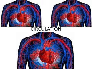
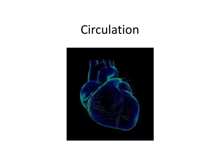
Circulation. The Importance of the Circulatory System. Delivers nutrients to the body. Composed of 96000km of blood vessels Delivers O 2 to 60 trillion cells Five litres of blood/min travels from heart to lungs. Carries wastes away from cells CO 2 is delivered to the lungs and exhaled

E N D
The Importance of the Circulatory System • Delivers nutrients to the body. • Composed of 96000km of blood vessels • Delivers O2 to 60 trillion cells • Five litres of blood/min travels from heart to lungs
Carries wastes away from cells • CO2 is delivered to the lungs and exhaled • Urine, ammonia is brought to the kidneys and excreted
Defense against invading organisms • Your immune system
Structures of the Circulatory System • Arteries • Carry blood away from the heart • Have a 3 layer structure. Middle layer is muscle which provides flexibility and stretch
Capillaries • Single layer of cells that form thin tubes. • Site of fluid and gas exchange • All cells of your body lie very near a capillary
Veins • Carry deoxygenated blood that has travelled through arteries and capillaries. • Returns the blood to the heart and lungs to be re-oxygenated • Contraction of skeletal muscles aids in moving blood against gravity
Circulation Pathway • Arteries carry blood away from heart • Blood passes into smaller arteries called arterioles • Blood is directed into capillaries. Site of gas exchange • Capillaries merge and become larger vessels called venules. • Venules merge to become veins.
The Heart • Is surrounded by a fluid filled membrane called the pericardium • The heart is not a single pump but 2 parallel pumps separated by a wall of muscle called a septum • Muscles contractions on the right side mirror contractions on the left side.
Heart Function • The pump on the right side receives deoxygenated blood from the body tissues and pumps it to the lungs • The pump on the left receives oxygenated blood from the lungs and pumps it to the cells of the body
Heart Structure • The human heart has 4 chambers. • 2 thin walled atria and 2 thick walled ventricles.
Atria • Receive blood from veins. • Right atrium receives blood from the superior and inferior vena cavae • Left atrium receives blood from the right and left pulmonary veins.
Ventricles • Pump blood through arteries • The right ventricle sends blood to the lungs via the pulmonary arteries • The left ventricle sends blood to the body via the aorta.
Other heart parts • Coronary arteries- supply the cardiac muscle with oxygen and nutrients • Semilunar valves- valves that prevent backflow from the arteries to the ventricles. • Atrioventricular valves- prevent backflow of blood from ventricles into the atria.
Review Questions • What are the 3 main functions of your circulatory system? • Name the 4 chambers of the mammalian heart. • What is the function of the valves in the heart? How many valves does the heart have? • Which blood vessel carries blood towards the heart? Away? • Briefly compare the structures of the 3 types of blood vessels and explain how their structures suits their function.
IN #3 • Draw a mammalian heart. Label major structures. Trace the flow of blood through the heart using red and blue pens.
The Heart Beat • The familiar “lubb-dubb” heart sounds are caused by the closing of the heart valves.
The “lubb” • Contraction of atrial walls forces blood through AV valves into ventricles. • Atria fill with blood as they relax Diastole (the lowest blood pressure before the ventricles contract. • As the ventricles begin to contract blood is forced up the sides of the ventricles and the AV valves close lubb
The “dubb” • Ventricular contraction increases pressure in the chambers, forcing blood through the semi lunar valves and out of the arteries Systole (the maximum pressure during the ventricular contraction) • Blood is prevented from re-entering the ventricles by the closing of the semi lunar valves dubb
Blood Pressure • As blood passes through the vessels in the body it exerts pressure against the vessel walls Blood Pressure • BP is measured in mmHg (millimeters of mercury) with a sphygmomanometer • It is presented as systolic pressure over diastolic pressure ex: 120/80 mmHg
What causes high blood pressure? • Health risk associated with high blood pressure?
Setting the heart’s tempo • Cardiac tissue is myogenic tissue. It contracts without external stimulation • The SA (sinoatrial) node regulates and coordinates the beating of all heart cells. • It is located where the vena cavae enter the right atrium
Acts as a pace maker and sets the rhythm at about 70 beats/min • The contractions travel to a second node the AV node. (atrioventricular) • AV node passes nerve impulses through the septum to the ventricles
Electrocardiograph is an instrument that monitors electrical activity of the heart • P wave atrial contraction • Q,R, S waves ventricular contractions • T wave ventrical recovery
Review Qs 1. Arrange the following structures in the order that blood flowing through the coronary pathway encounters them: pulmonary artery, pulmonary vein, aorta, superior vena cava, right atrium, left atrium, right ventricle, left ventricle. Begin with the pulmonary artery. 2. Describe the role of the AV and SA nodes in setting the heart’s tempo.
IN #4 • Using a flow chart or other graphic organizer illustrate how the “lubb” and the “dubb” sounds of the heart are created. (If you use a diagram be sure to label it. )