Download

1 / 49
541 likes | 937 Views
The control of the heart rate. Cerebral cortex. The hypothalamus and limbic system. Inspiratory center. Cardiovascular centers. Chemical factors Physical factors Mechanical factors. S-A node. Reflexes. The peripheral resistance.

E N D
The control of the heart rate Cerebral cortex The hypothalamus and limbic system Inspiratory center Cardiovascular centers Chemical factors Physical factors Mechanical factors S-A node Reflexes
The peripheral resistance As the blood flows from the arterial to the venous side of the circulation, it meets resistance because of the smaller caliber of the vessels and the viscous nature of the blood. This is called the peripheral resistance. It is an important factor in generating and maintaining the arterial blood pressure. Vasoconstriction of the small vessels increases the peripheral resistance, which in turn elevates the arterial blood pressure. Whilst vasodilatation decreases the resistance and lowers the pressure. The main factor is a gradient of blood pressure.
RESISTANCES IN SERIES RT = RA + RC + RV RESISTANCES IN PARALLEL R1 PV PA 1 RT 1 R1 1 R2 1 R3 R2 = + + R3 1 RT = 1 R1 1 R2 1 R3 + +
Pressure Drop in the Vascular System ELASTIC TISSUE MUSCLE LARGEARTERIES SMALLARTERIES MEANPRESSURE ARTERIOLES CAPILLARIES VENULES &VEINS LARGE SMALL LARGE INSIDE DIAMETER
Nervous factors The most important factor in the regulation of the heart rate is the activity of the cardiovascular centers in the medulla oblongata. This activity is transmitted to the heart via its sympathetic and parasympathetic nerve supply.
Sympathetic nerve supply There is a resting sympathetic tone that tends to increase the heart rate up to 120 beats/min. This tone is weak and is masked by the strong inhibitory vagal tone that decreases the heart rate down to 75 beats/min during rest. However, stimulation of the sympathetic cardiac nerves has a +ve chronotropic effect. The heart rate may go up to 200 beats/min. The sympathetic chemical transmitter noradrenaline decreases the permeability of the pacemaker membrane to K+. This accelerates the depolarization of the membrane → shortens the duration of the pacemaker potential → increases the frequency of discharge of impulses from the S-A node → increases the heart rate.
Parasympathetic nerve supply There is a resting inhibitory vagal tone that keeps the heart rate at its resting level of ~ 75 beats/min. During deep quite sleep, the vagal tone increase and the heart rate decreases down to 60 beats/min. Vagal stimulation has a –ve chronotropic effect. The parasympathetic chemical transmitter acetyl choline increases the permeability of the pacemaker membrane to K+. This slows down the depolarization of the membrane → prolongs the duration of the pacemaker potential → deccreases the frequency of discharge of impulses from the S-A node → decreases the heart rate.
Heart rate A change in the heart rate produces a stepwise change in the force of myocardial contraction until a final steady level of contractility is reached. has a negative inotropic The steady level of myocardial contractility is directly proportional to the heart rate, within limits. In other words, cardiac acceleration has a +ve inotropic effect and cardiac slowing effect.
INCREASING HEART RATE INCREASES CONTRACTILITY Ca++ Ca++ Normal Heart Rate Fast Heart Rate Ca++ Ca++ Ca++ Ca++
CARDIAC FUNCTION CURVE Cardiac Output = Stroke Volume x Heart Rate Constant If: STROKE VOLUME Then: CO reflects SV DIASTOLIC FILLING Right Atrial Pressure (RAP) reflects Diastolic Filling
CARDIAC FUNCTION CURVE THE FRANK- STARLING “LAW OF THE HEART” 15- 10- CARDIAC OUTPUT (L/min) Pressure 5- Volume -4 0 +4 +8 RAP mmHg
CARDIAC FUNCTION CURVE THE FRANK- STARLING “LAW OF THE HEART” 15- Increased Contractility 10- CARDIAC OUTPUT (L/min) 5- -4 0 +4 +8 RAP mmHg
CARDIAC FUNCTION CURVE THE FRANK- STARLING “LAW OF THE HEART” 15- 10- Decreased Contractility CARDIAC OUTPUT (L/min) 5- -4 0 +4 +8 RAP mmHg
CARDIAC FUNCTION CURVE THE FRANK- STARLING “LAW OF THE HEART” 15- Increased Heart Rate 10- CARDIAC OUTPUT (L/min) 5- -4 0 +4 +8 RAP mmHg
CARDIAC FUNCTION CURVE THE FRANK- STARLING “LAW OF THE HEART” 15- 10- Decreased Heart Rate CARDIAC OUTPUT (L/min) 5- -4 0 +4 +8 RAP mmHg
CARDIAC CENTRES & CARDIAC INNERVATION • Outline: • Cardiac Centers: • - Pressor Area = vasomotor area or vasomotor centre • (VMC) • - Depressor Area = cardiac inhibitory centre (CIC) • Cardiac Innervations: • - Sympathetic nerve supply • - Parasympathetic nerve supply • Arterial baroreceptors and peripheral chemoreceptors • Further Reading: • Guyton: Textbook of Medical Physiology • Ganong: Review of Medical Physiology
HEART RATE & ITS REGULATION • CARDIAC CENTRES AND CARDIAC INNERVATION: • The activity of the heart (CVS) is under the control of 2 bilateral areas in the medulla oblongata: Pressor area and depressor area. • THE PRESSOR AREA: • - It is also called the vasomotor area or vasomotor centre (VMC). • - It is present in the ventrolateral parts of the medulla oblongata and it is connected with preyganglionic sympathetic neurons in the spinal cord. • - The Pressor area contains 2 centers: • a) Cardiac acceleratory centre (CAC); also called cardiac stimulatory centre (CSC). • b) Vasoconstrictor centre (VCC)
Stimulation of the Pressor area produces sympathetic effects i.e. • a) Increase of heart rate and increase of myocardial • contractility • b) Vasoconstriction of the arterioles • Normally and under resting condition, the VCC discharges impulse continuously at a certain rate. This is called vasomotor tome (= vasoconstrictor sympathetic tome) which leads to partial VC of the arterioles and venules all over the body. • of the VM tone more vasoconstriction • of the VM tone less vasoconstriction (=vasodilatation) • THE DEPRESSOR AREA: • - It is inhibitory area in the medulla oblongata and it • contains a cardio-inhibitory centre (CIC) [=dorsal • motor nucleus of the vagus nerve].
- Stimulation of this area produces parasympathetic (vagal) effects on the heart i.e. decrease of heart rate and decrease of atrial contractility. • - Normally and under resting condition; the CIC discharges continuous inhibitory impulses along the vagus nerve to the heart. This is called vagal tone which checks the high inherent rhythm of the SA node. • of vagal tone to the heart of heart rate • of vagal tone to the heart of heart rate • INNERVATION OF THE HEART: • - The heart receives its nerve supply from both divisions • of the ANS i.e. • Sympathetic nervous system and • Parasympathetic nervous system
CARDIOVASCULAR CENTRES (CVCs) • - CARDIOVASCULAR CENTRES are present in the medulla oblongata in 2 areas: • 1) Pressor area: which contains CAC (CSC) and VCC • 2) Depressor area: which contains CIC & VOC • THE PRESSOR AREA: • - It contains 2 centers: • 1) CAC = cardiac accelerator centre • = CSC= cardiac stimulatory centre • 2) VCC = vasoconstrictor centre • - Stimulation of Pressor area sympathetic effects: • 1) heart rate • 2) VC of the arterioles and venules
Normally, during rest the VCC discharges continuously at a certain rate i.e it exerts a tone known as vasoconstrictor tone (sympathetic tone) partial VC of the arterioles. • DEPRESSOR AREA: • - It contains • CIC = Cardiac Inhibitory Centre • - Stimulation of the depressor area parasympathetic effects: • heart rate. • - Normally, during rest the CIC discharges continuously at a certain rate through the vagus nerves i.e. it exerts a tone known as vagal tone (parasympathetic tone) HR.
SYMPATHETIC NERVE SUPPLY: • - The preyganglionic sympathetic fibers arise from the lateral horn cells of the upper 4 thoracic segments of the spinal cord (T1-T4). • - The preyganglionic fibers relay in the cervical ganglia (superior, middle & inferior) and the upper 4 thoracic ganglia of the sympathetic chain. • - Postganglionic fibers arise from these ganglia to supply: • The atria and the ventricles of the heart including the specialized tissues (SA node, AV node, AV bundle, • bundle branches and the purkinje fibers) • The coronary vessels • - FUNCTIONS OF SYMPATHETIC CARDIAC NERVES: • 1) Stimulation of all properties of the cardiac muscle • 2) Vasodilatation of the coronary arteries • 3) Increase of O2 consumption of the cardiac muscle
PARASYMPATHETIC NERVE SUPPLY: • - The parasympathetic supply is through the two vagi • - The preyganglionic vagal fibers arise from the dorsal vagal nucleus (CIC) in the medulla oblongata. • - The preyganglionic fibers relay in terminal ganglia located in the atria • - The postganglionic fibers are short; they arise from the terminal ganglia to supply the atrial muscle, SA node, AV node, main stem of the AV bundle and the coronary vessels. • - FUNCTIONS OF THE PARASYMPATHETIC SUPPLY: • 1) Inhibition of all properties of the cardiac muscle • Stimulation of all properties of the cardiac muscle • 2) Vasoconstriction of the coronary arteries • 3) Decrease of O2 consumption of the heart
VAGAL TONE: • Vagal Tone is the continuous inhibitory impulses carried by the vagus nerve from the CIC to the heart to inhibit the high inherent rhythm of the SA node. This occurs under resting condition and produces a basal heart rate (about 70/ min). • Vagal tone is a baroreceptors reflex i.e. it is produced by impulses from the baroreceptors present in the aortic arch and carotid sinus. These impulses stimulate the CIC. • Evidences of Vagal tone: • 1) Injection of atropine (=parasympathetic drug) causes increase of heart rate. • 2) Cutting of both vagi in experimental animals causes increase of heart rate. • At rest, the vagal tone to the heart is dominant over the weak sympathetic tone. During muscular exercise, heart rate is increased due to decrease of vagal tone and increase of sympathetic activity.
THE CARDIOVASCULAR RECEPTORS: • The walls of the heart and some blood vessels contain specific types of sensory receptors for several reflexes which control and circulation and respiration. • Examples: • Arterial baroreceptors and peripheral chemoreceptors • Atrial receptors • Ventricular receptors • Pulmonary receptors • The most important of these receptors are: • 1) The arterial baroreceptors located in the aortic arch and carotid sinus • 2) The peripheral chemoreceptors located in the aortic and carotid bodies • 3) The atrial (volume or stretch) receptors located in the • right atrium
THE ARTERIAL BARORECEPTORS OF THE AORTIC ARCH AND CAROTID SINUS: • These receptors are stretch receptors located in the wall (adventia) of • the aortic arch (=curve between the ascending and descending parts of the aorta). • the carotid sinus (= dilation at the beginning of the internal carotid artery. • These receptors send their afferent impulses through 2 nerves: • the aortic nerve which is a branch of the vagus nerve (10th cranial nerve) • the sinus nerve which is a branch of the • glassopharyngeal nerve (=9th cranial nerve)
THE TWO NERVES ARE CALLED THE BUFFER NERVES • The arterial baroreceptors are not stimulated at all by arterial pressures between 0 and 60 mm Hg; but above 60 mm Hg they start to discharge impulses to the cardiovascular centers in the medulla oblongata along the buffer nerves. • The rate of discharge from the baroreceptors is directly proportional to the systemic ABP i.e. the higher the blood pressure, the higher the frequency of impulses generated in the baroreceptors. • The maximal discharge from the baroreceptors occurs at arterial blood pressure of about 180 mm Hg (180-200 mm Hg).
Functional of the baroreceptors “the baroreceptorsreflexes”: • The arterial baroreceptors are sensitive to any change in the ABP, so they are important to keep the ABP normal (through baroreceptors reflexes) • At normal level of ABP, the baroreceptors discharge excitatory impulses to the depressor area (CIC) and inhibitory impulses to the Pressor area (VMC or CAC & VCC) at a certain rate • - Stimulation of CIC which produces normal vagal tone (resting heart rate) • - Inhibition of CAC • - Inhibition of the inherent high activity of the VCC • partial VC.
Therefore, at normal ABP, the baroreceptors discharge • normal degree of vagal tone (=basal heart rate) and • sympathetic vasoconstrictor tone (=partial VC of the • arterioles). • When the ABP is increased, the rate of discharge from the baroreceptors to the medullar CV centers is also increased • More stimulation of the depressor area (CIC) increase of vagal tone and decrease of heart rate. • More inhibition of the Pressor area (=VMC = VCC) vasodilatation of the arterioles. These effects (HR + VD) may decrease the high BP towards normal.
When the ABP is decreased, the rate of discharge from the baroreceptors to the medullar CV centers is also decreased • Inhibition of the depressor area (CIC) decrease of vagal tone and increase of heart rate. • Stimulation of the vasomotor centre (Pressor area) marked vasoconstriction dilatation of the arterioles. These effects (HR + VD) may decrease the high BP towards normal. The arterial baroreceptors of the aortic arch and carotid sinus; their afferent connections to the medullar CV centers and the efferent pathways from these centers to the heart and the arterioles constitute a reflex feedback control mechanism that operates to stabilize the ABP i.e:
The arterial baroreceptors reflex mechanism: • Feedback control system for regulation of ABP • Arterial pressure buffer system (i.e. buffers acute changes in ABP). • Moderator mechanism (i.e. it moderates acute changes in ABP). THE PERIPHERAL CHEMORECEPTORS OF THE AORTIC & CAROTID BODIES • The peripheral chemoreceptors are located in • The aortic body which lies very close to the aortic arch • The carotid body which lies very close to the carotid sinus. • These receptors have rich blood supply i.e. they have high rate of blood flow in relation to their size.
FUNCTION OF THE PERIPHERAL CHEMORECEPTORS “ the chemoreceptor reflexes”: • The peripheral chemoreceptors are sensitive to changes in H+ concentration (pH). • If PO2, PCO2 & pH are normal in the arterial blood, these receptors send impulses (at a certain rate) along the buffer nerves to CV centers in the medulla oblongata - Inhibition of the depressor area (CIC). - Stimulation of the Pressor area (VMC) partial VC of the arterioles • If PO2 is decreased (hypoxia), PCO2 is increased (hypercapnia) , or H+ conc. is increased (= pH or acidosis), the peripheral chemoreceptors are stimulated and they discharge more impulses to the medullar CV centers - More inhibition of the depressor area (CIC).
- More stimulation of the Pressor area (VMC) increase of the Pressor area (VMC) increase of heart rate and vasoconstriction of the arterioles increase of ABP. This “chemoreceptor” reflex occurs in case of acute drop of the ABP to 40-60 mm Hg as during severe haemorrhage. This is because of the rich blood supply of the peripheral chemoreceptors which makes them sensitive to changes in ABP. Thus, ABP ischemia of these receptors local hypoxia (O2 lack) their stimulation which in turn, excites the vasomotor area HR & VC ABP towards normal. • N.B: • Central chemoreceptors are present in the medulla oblongata and they are sensitive to H+ changes in the cerebrospinal fluid (CSF). • The baroreceptors are more concerned with regulation of circulation and the chemoreceptors are more concerned with regulation of respiration.
REGULATION OF HEART RATE • Outline: • Normal value and methods of counting of heart rate (HR) • Physiological variations of heart rate • Nervous regulation of heart rate (HR): • - Bainbridge reflex, Mary's reflex (law) & respiratory • sinus arrhythmia • - Alam-Smirk reflex and trigger Jones reflexes. • Chemical regulation of HR (effect of hypoxia, hypercapnia, hormones & drugs). • Physical regulation of HR (effect of hyperthermia & hypothermia). • Tachycardia and bradycardia: causes of exercise tachycardia • Further Reading: • Guyton: Textbook of Medical Physiology • Ganong: Review of Medical Physiology
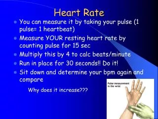
REGULATION OF HEART RATE • The normal heart rate (=number of heart beats/ min) is about 70 minute. The heart rate can be counted by: a) Palpitation of the arterial pulse (e.g. radial pulse) or palpitation of the apex. b) Auscultation of the heart sounds c) ECG (=electrocardiogram) • The resting heart rate is determined by the degree of the vagal tone i.e. increase if vagal tone decrease of heart rate & decrease of vagal tone increase of heart rate.
The resting heart rate is determined by the degree of the vagal tone i.e. increase if vagal tone decrease of heart rate & decrease of vagal tone increase of heart rate. Vagal tone is greater in males than females, in adults than in children and athletes than non-trained persons. Therefore, physiological variations in heart rate are related to age, sex, physical training and metabolic rate. • Regulation of heart rate includes 3 mechanisms: a) Nervous regulation: Changes in heart rate by afferent impulses that modify the activity of the cardiac centers in the medulla oblongata. b) Chemical regulation: Changes in heart rate due to changes in the chemical composition of blood. c) Physical regulation: Changes in heart rate due to changes in body (blood) temperature.
(a) NERVOUS REGULATION • Nervous regulation of heart rate depends on afferent impulses that reach the cardiac centers in the medulla oblongata to change their activity changes on the heart rate. 1) Impulses from the right atrial receptors “Bainbridge reflex”: - Bainbridge reflex “is the reflex increase of heart rate due to increase of the right atrial pressure”. Therefore, increase of venous return and venous pressure in the right atrium (e.g. during muscular exercise) causes reflex heart acceleration. - The increased right atrial pressure stimulation of stretch receptors (=volume receptors) in the atrial wall discharge of impulses along afferent vagal fibers to the medulla oblongata stimulation of the vasomotor centre
efferent impulses along the sympathetic nerves to the heart increase of the heart rate. - Cardiac acceleration helps pumping of excess venous return into the arterial side of the circulation, so it prevents stay nation of blood in veins. • Impulses from the arterial baroreceptors of the aortic arch & carotid sinus “Mary's reflex”. - Mary's reflex (Mary's Law) states that “the heart rate is inversely proportional to the arterial blood pressure “ provided that other factors affecting heart rate remain constant. Thus, increase of ABP decrease of heart rate & decrease of ABP increase of heart rate.
- Marey’s reflex is a baroreceptors reflex i.e. • ABP stimulation of the arterial baroreceptors in the aortic arch and carotid sinus afferent impulses along the buffer nerves stimulation of cardio inhibitory centre (CIC) vagal tone and in turn decrease of heart rate. • ABP (as in haemorrhage) decrease of number of impulses from the arterial baroreceptors to the CV centers in the medulla oblongata inhibition of the CIC and stimulation of the vasomotor centre (VMC) increase of heart rate. • Impulses from the respiratory centre and the lungs: “Respiratory sinus arrhythmia” • Normally, there is regular increase of heart rate during inspiration and decrease of heart rate during expiration. This phenomenon is called respiratory sinus arrhythmia. It occur during deep respiration.
The increase of heart rate during inspiration may be due to inhibition of the depressor area (CIC) and decrease of the vagal tone by the following mechanisms: • a) During inspiration, the activity of the inspiratory centre irradiates inhibitory impulses to CIC. • b) During inspiration, expansion of the lungs stimulation of stretch receptors in the wall of the alveoli discharge of impulses along afferent pulmonary vagal fibers inhibition of CIC. • c) During inspiration, the venous return to the heart is increased stimulation of the stretch receptors in the right atrium discharge of impulses along afferent vagal fibers inhibition of CIC.
Impulses from the higher centers (cerebral cortex & hypothalamus): • Certain areas in the cerebral cortex can influence heart rate through their effects on the hypothalamus and the cardiac centers in the medulla oblongata e.g. • During emotions & muscular exercise, impulses from the cerebral cortex stimulation of the vasomotor centre increase of heart rate. • The conditioned reflexes which mediated via the cerebral cortex increase or decrease of heart rate in response to visual or auditory stimuli. • The hypothalamus also contain nuclei which can modify heart rate e.g. during sleep or emotions.
Impulses from other parts of the body: • a) Skeletal muscles (Alam – Smirk reflex): • - During muscular activity, the proprioceptors of the active muscles discharges impulses along afferent nerve fibers to the medulla oblongata stimulation of the vasomotor centre (VMC) increase of heart rate to supply the active muscles with more blood. • b) Trigger areas (eyeball, ear, larynx, epigastrium, testicles… etc): • - If painful stimuli (e.g. heavy blows) are applied to one of the trigger areas, this leads to reflex decrease of heart rate (bradycardia). • Slight or moderate (sematic or visceral) pain usually causes increase of heart rate. However, severe pain (specially visceral pain) is usually associated with decrease of heart rate.
(b) CHEMICAL REGULATION • This includes the effect of changes in blood gases (O2 and CO2), the effect of some hormones (thyroxin, adrenaline & noradrenalin) and the effect of some autonomic drugs (e.g. adrenaline & atropine). • Effect of changes in PO2 and PCO2: This includes the effect of changes in blood gases (O2 and CO2), the effect of some hormones (thyroxin, adrenaline & noradrenalin) and the effect of some autonomic drugs (e.g. adrenaline & atropine). • Hypoxia (O2 Lack): - Slight or moderate hypoxia PO2 in blood increase of heart rate due to stimulation of the peripheral chemoreceptors in the aortic and carotid bodies stimulation of the vasomotor centre in the medulla oblongata = “chemoreceptor reflex”.
- hypoxia occurs in anemia, heart failure, haemorrhage and at high attitudes. - Severe hypoxia decrease of heart rate (brady cardia) due to direct depression of the SA node. • Hypercapnia (increased CO2): - Slight or moderate hypercapnia PCO2 in blood increase of heart rate due to: - Direct stimulation of the vasomotor centre in the medulla oblongata. - Stimulation of the peripheral chemoreceptors in the aortic and carotid bodies stimulation of the vasomotor centre (=chemoreceptor reflex”. • Severe Hypercapnia (=marked Co2 excess in blood) decrease of heart rate due to direct depression of the SA node.
Effect of hormones (thyroxin, adrenaline & noradrenalin): • Thyroxin: - Thyroxin increases heart rate due to a) Direct stimulation of the SA node and increase of its sensitivity to catecholamine. b) Increase of metabolic rate. • Adrenaline: - Adrenaline (like sympathetic) causes increase of heart rate due to direct stimulation of the SA node. • Noradrenalin: - Noradrenalin is a strong vasoconstrictor agent generalized vasomotor constriction ABP. Increase of ABP decrease of heart rate (Marey’s Reflex). • Effect of autonomic drugs: • Parasympatholytic drugs (e.g. atropine) increase of heart rate. • Sympathomimetic drugs (e.g. adrenaline) increase of heart rate.
(c) PHYSICAL REGULATION Effect of changes in the blood (body) temperature: • Increase of blood temperature (hyperthermia or fever): - Increase of the blood temperature above normal increase of heart rate due to: a) Direct stimulation of the SA node. b) Stimulation of the vasomotor centre in the medulla oblongata by impulses discharged by the hypothalamus (thermo-regulatory centre). - Arise of 1°C in the blood (body) temperature increase of heart rate by about 10 beats. However, in diphtheria, the heart rate is decreased though the body temperature is increased. This is due to the effect of diphtheria toxins on the heart depression of the cardiac muscle.
Decrease of blood temperature (hypothermia): - Decrease of the blood (body) temperature below normal brady cardia TACHYCARDIA & BRADYCARDIA • Tachycardia means increase of heart rate. It may be physiological or pathological: • Physiological e.g. as during emotions and muscular exercise. • Pathological e.g. as in fevers, hyperthyroidism & haemorrhage. • Bradycardia means decrease of heart rate. It may be physiological or pathological: • Physiological as during quiet sleep and well trained athletes (due to high vagal tone). • Pathological as in hyperthermia, hypothyroidism & heart block
Causes of heart acceleration during muscular exercise: • Heart rate is markedly increases (140/min or more) during muscular exercise. This is due to: 1) Emotional Effect by impulses from the cerebral cortex and hypothalamus stimulation of the vasomotor centre. 2) Chemoreceptor reflex i.e. stimulation of the peripheral chemoreceptors in the aortic and carotid bodies by PO2 & PCO2 + H+ 3) Bainbridge Reflex i.e. due to increase of venous pressure in the right atrium which results from increase of venous return. 4) Reflex activation of the vasomotor centre by afferent impulses from the proprioceptors of the active muscles. • 5) Secretion of adrenaline from the adrenal medulla • direct stimulation of the SA node. • 6) Sympathetic over activity stimulation of • sympathetic nerves of the heart. • 7) Increase of the blood temperature during exercise stimulation of the SA node.