Download
1 / 59
650 likes | 1.29k Views
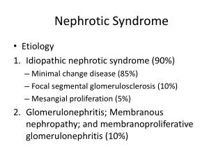
Secondary Causes of the Nephrotic Syndrome. Sumit Kumar, MD Presbyterian Hospital Dallas Dallas, TX. Normal Anatomy. Classification of Glomerular Diseases. Primary glomerular diseases Nephrotic Syndrome Non Immune Complex Immune Complex Nephritic syndrome

E N D
Secondary Causes of the Nephrotic Syndrome Sumit Kumar, MD Presbyterian Hospital Dallas Dallas, TX
Classification of Glomerular Diseases • Primary glomerular diseases • Nephrotic Syndrome • Non Immune Complex • Immune Complex • Nephritic syndrome • Post infectious glomerulonephritis • Ig A nephropathy / Henoch Schonlein purpura
Classification of Glomerular Diseases • Diseases Associated With Nephrotic Syndrome • Monoclonal Immunoglobulin Deposition Disease (MIDD) • Amyloidosis • Light chain deposition disease • Others • Infections • Deposition diseases • Secondary FSGS • Malignancies
Classification of Glomerular Diseases • Diseases Associated With Nephritic ± Nephrotic Syndrome or RPGN • Immune Mediated: Lupus; cryoglobulin related; anti-GBM • Pauci immune
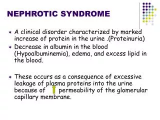
Proteinuria • Proteinuria>150mg/24hr • Dipstik Test: • >1(+) • False positive: gross hematuria • Urine protein:urine creatinine: • Normal <0.15 • Ratio of 1 correlates with proteinuria of 1g/24hr • Most accurate is 24 hr urine collection for protein and creatinine • Pitfalls of spot protein testing: False positives and negatives
Types of Proteinuria • Glomerular • Primary and Secondary Diseases • Charge selectivity (as in MCD) • Hemodynamic (severe HTN and CHF) • Tubular • Fanconi Syndrome, Tubulo-interstitial disease • Overflow • Monoclonal gammopathies • Inflammatory • Variants • Transient: Usually disappears, No workup needed • Orthostatic • Associated with exercise, fever, stress
Clinical Pathways for the Pathogenesis of Glomerular disease Asymptomatic hematuria Macroscopic Hematuria Nephritic Syndrome Nephrotic Syndrome RPGN Glomerular Disease Chronic Glomerulonephritis ESRD
Hereditary:Rare Infectious: Viral: HBV, HCV, HIV Bacterial: 2 syphilis, SBE Protozoan: Ch malaria Immunologic:SLE Drugs: Gold, Penicillamine, NSAIDs Metabolic: Diabetes Mellitus Amyloidosis Neoplasms: Myeloma Solid e.g. colon, lung, breast Lymphoma, leukemia Miscellaneous: Massive obesity, sleep apnea syndrome, chronic reflux nephropathy (FSGS) Secondary Causes of the Nephrotic Syndrome
Secondary Glomerular Diseases • Diabetic Nephropathy • Lupus Nephritis • Secondary focal segmental glomerulosclerosis • Secondary membranous glomerulopathy • Membranoproliferative Glomerulonephritis • Paraproteinemia • Collagen Vascular Disease • Malignancy associated
Doc…..I think I am not well!! • Periorbital edema • Pedal edema • Weight gain
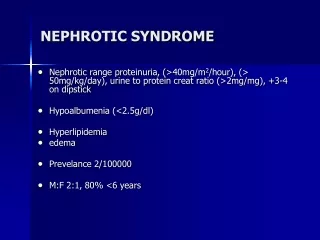
Approach to the Patient with Nephrotic Syndrome • Make the diagnosis Proteinuria > 3.5 g/1.73 m2 Hypoalbuminemia Edema Lipuria Hypercholesterolemia • Screen the patient for secondary etiologies
Assessment of a Nephrotic patient • History: medications, drugs, surgeries, infections, obesity, travel, family history • Serologies • Hepatitis B and C, HIV • ANA, C3, C4, SPEP, UPEP, Cryoglobulins • VDRL, ASO titer • Imaging • Chest Xray • Renal sonogram
Prevalent counts & adjusted rates, by primary diagnosis Point prevalent ESRD patients; Medical Evidence form data; rates adjusted for age, gender, & race.
Projected growth of the U.S. diabetic population, by race White Black Other 1978 4,325,230 824,354 117,175 1990 6,475,204 1,319,594 385,756 2000 8,786,661 2,005,805 773,134 2030 18,441,647 5,539,283 2,941,056 Prevalence rates of diabetes in 1980–1998 obtained from National Health Interview Survey (NHIS) data. These data are linearly extrapolated to obtain prevalent rates for 1978–2030, & estimates are then multiplied by census projections to obtain estimated numbers of individuals with diabetes for 1978–2030.
The Natural History of Diabetic Nephropathy in IDDM Incipient NephropathyHyperfiltration Blood Pressure Poor glycemic control Onset of Hypertension 15-40 0 2 5 10 -30 13-25 Time (yrs) Onset of Diabetes Onset of Proteinuria GFR ESRD Functional Changes GFR Reversible albuminuria Kidney size Structural ChangesGBM thickening Mesangial expansion
Natural History of Diabetic Nephropathy in NIDDM- Lessons learnt from the Pima Indians • Nelson et al NEJM 1996;335:1636-1642
Natural History of Diabetic Nephropathy in NIDDM- Lessons learnt from the Pima Indians • Nelson et al NEJM 1996;335:1636-1642
Major Therapeutic Maneuvers to Slow Loss of GFR in Diabetic Nephropathy Normotension Hyperglycemia Euglycemia Protein restriction ACEi, ARB Lipid Management Weight loss, exercise, smoking cessation Glomerulosclerosis
Lupus Nephritis • Renal involvement • Early Course: 30-50% of unselected patients • Later Course: 60-80% • Most patients present with proteinuria • Hypertension ± • Hyperkalemic renal tubular acidosis
Lab tests in Lupus Nephritis • ANA, Anti DNA antibody, Anti Smith antibody • Anemia of moderate degree, Coombs + in minority, severe hemolytic anemia – rarely • Leukopenia, thrombocytosis • Hypocomplementemia: C4 and C1q are depressed more than C3 suggesting classic pathway activation (never occurs in idiopathic MPGN • Antiphospholipid antibody - ⅓ - ½of patients with lupus nephritis • Renal arterial, venous and glomerular cap thrombosis, Libman-Sacks arthritis, cerebral thrombosis
Diagnosis and Differential Diagnosis • Suspect in • Middle aged, nephrotic male • Idiopathic membranous nephropathy in a young woman • Routine screening of all nephrotic patients with ANA • Differential: Rheumatoid arthritis; Henoch Schonlein purpura; Ig A nephropathy; vasculitis
WHO Class II Lupus Nephritis • Mesangial disease – 10-25% of all biopsies • Mesangial expansion • Clinically mild disease – non nephrotic proteinuria; normal renal function
WHO Class III Lupus Nephritis • Focal Proliferative • 20-35% of biopsies • Focal necrosis • Clinically mild disease – proteinuria; hematuria ±
WHO Class IV Lupus Nephritis • Diffuse proliferative (DPGN) • 35-60% of biopsies • Hypercellularity • Intense inflammation • Clinically – hematuria, red cell casts, proteinuria, hypertension, acute renal failure. • Most amenable to treatment • Rx: NIH Protocol
WHO Class V Lupus Nephritis • Membranous • 10-15% of biopsies • Silver positive “spikes” – subepithelial • Clinically nephrotic, often normal renal function • Rx: Difficult to treat; Steroids; Ponticelli protocol
Severe Nephrotic Syndrome Drugs: heroin; NSAIDs Viruses: Hep B, HIV, parvo Non Nephrotic proteinuria Reduced Mass Solitary kidney, allograft, surgical ablation, renal dysplasia, agenesis, segmental hypoplasia Non Nephrotic Proteinuria Scarring VUR, hypertensive nephrosclerosis, post infectious or inflammatory; lupus nephritis; vasculitides Hyperfiltration Obesity, sickle cell nephropathy, congenital cyanotic heart disease Other Causes Malignancies: lymphomas Misc: sarcoidosis, radiation nephritis; Charcot-Marie-Tooth Classification of Secondary FSGS
Secondary FSGS • HIV associated Nephropathy (HIVAN) • Vesicoureteric reflux • Obesity
HIV associated Nephropathy • Massive proteinuria • Micro-hematuria • Azotemia • Rapid progression to ESRD • African American Patients • CD4 count low • Normotensive • Sonogram
HIV associated Nephropathy • Natural History • Malignant course • ESRD within 3 to 4 months • Less common now than 10 years ago • Differential Diagnosis • Heroin associated Nephropathy • Options • Dialysis • HIV treatment • Transplantation??
Secondary FSGS • Vesicoureteric reflux • May not occur for several years • Often a poor prognostic sign – strong correlation between extent of glomerular involvement and the magnitude of proteinuria and GFR decline\ • Hypertension occurs late • Histology: Hyalinosis in unscarred areas of the kidney or in the contralateral normal kidney
Secondary FSGS • Obesity • Proteinuria is fairly common (upto 40%) • Likely to be part of the endothelial dysfunction syndrome a.k.a. dysmetabolic syndrome X • Remission of proteinuria often occurs with weight reduction • Association with sleep apnea
Case Presentation - Amyloidosis HOPI • B.J.A., a 56 AA female with a long standing h/o HTN for the past 30 years was in good health until about 8 weeks prior to presentation. • Insidious onset of fatigue; 5 kg lost over a 2 m period. She had a baseline Cr of 1.6 three m prior. • About a week prior, she was started on Atenolol and HCTZ for recent difficulty in controlling her BP. • at that time, a Captopril renography was performed after Captopril 25 mg was given - Study was negative. • Over the course of the next week, her symptoms of fatigue worsened and she was admitted to DGH with progressively worsening SOB, 3-4 pillow orthopnea, and 2 episodes of syncope. • Hypertension for 30 yrs • Nifedipine XL 90 mg po qd, HCTZ 25 po qd, Atenolol 25 mg po qd and ASA PMH Meds
Case Presentation - Amyloidosis • 94/56; Wt 54 kg nd Ht 5’2” Eyes revealed Gr II HTNsive retinopathy JVP : 13 cm Chest had rales on the bases Precordial heave and a PSM was heard at the apex 3/6 rad to axilla Liver was 3cm BCM, Firm and non tender • Urine: SG 1.020, Protein 2+, Normal sediment Chem-7: Na 135, K 4.3, Cl 96, HCO3, BUN 76 and Cr 6.7 • CXRMild Pulmonary edema • EKG: NSR, LVE, LAFB PE Labs