Download
1 / 17
180 likes | 373 Views
Muscle Lab. Seminar Notes. Muscle Contraction. Sequence of events in muscle contraction AP in cell membrane AP travels down T-tubules Excitation-Contraction Coupling to sarcoplasmic reticulum Release of calcium Interaction of calcium with troponin Troponin interacts with tropomyosin

E N D
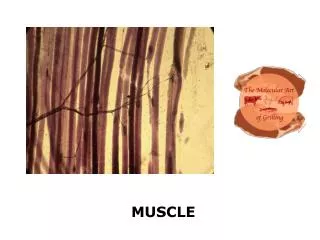
Muscle Lab Seminar Notes
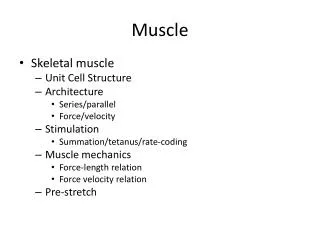
Muscle Contraction • Sequence of events in muscle contraction • AP in cell membrane • AP travels down T-tubules • Excitation-Contraction Coupling to sarcoplasmic reticulum • Release of calcium • Interaction of calcium with troponin • Troponin interacts with tropomyosin • Tropomyosin moves and myosin binding sites on actin are uncovered • Actin and myosin form crossbridges
Muscle Contractions • With multiple stimuli the fiber (or whole muscle) exhibits summation -tetany -fatigue. • Summation is a result of the fact that the second stimulus appears BEFORE all the calcium has returned to the SR. • Why is the threshold, measured by stimulating the nerve, LESS than the threshold for stimulating the muscle DIRECTLY? • In the first case, less stimulus energy is needed to bring the entire tissue (the nerve) to threshold and the fact that the nerve branches within the muscle.
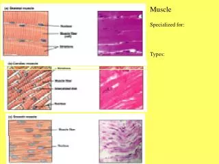
Electromyography Experiments • The electrical signal recorded from a contracting muscle is called an electromyogram. The EMG activity is a result of the electrical (ionic) activity in the many muscle cells (fibers) that make up a gross anatomical muscle
Voluntary Change in Force • Increase in EMG amplitude with increased load • books
Alternating Activity and Coactivation • Recording from the triceps and biceps • Note that when the biceps muscle is forcefully activated, the triceps activity is also increased. This is coactivation. • The effect is to stabilize the joint as the biceps produces the major force of commanded movement
Evoked EMG • Evoked EMG by stimulation of the median nerve • Increased amplitude of EMG as a function of increased stimulus • Also observe latency between stimulus and contraction
Conduction Velocity • Calculating the conduction velocity requires two bits of information • Distance and time • Distance from point of stimulus to recording site of EMG • Time it takes to travel from the stimulus site to the recording site
Stimulation of Median Nerve at Elbow to Determine Conduction Velocity
Conduction Velocity • Range = 1 – 120 m/s • Influenced by: Diameter of fiber Temperature Myelinated vs. non-myelinated
Muscle Experiments • Effects of nerve stimulation • Ulnar nerve • At wrist • At elbow • Observation of muscle movements
Twitch Response and Recruitment • Measurement of muscle contraction using a transducer • Muscle twitch • Single contraction and relaxation as a result of single stimulus • Recruitment • Increase in the amplitude of the twitch as a result of an increase in the amplitude of the stimulus • More fibers are brought into the response
MUSCLE TWITCH With a single stimulus a muscle fiber (cell) exhibits an all-or-none response. It contracts or it doesn't.