Download
1 / 28
350 likes | 951 Views
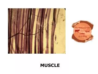
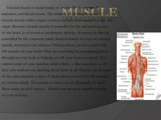
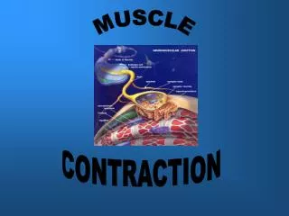
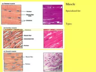
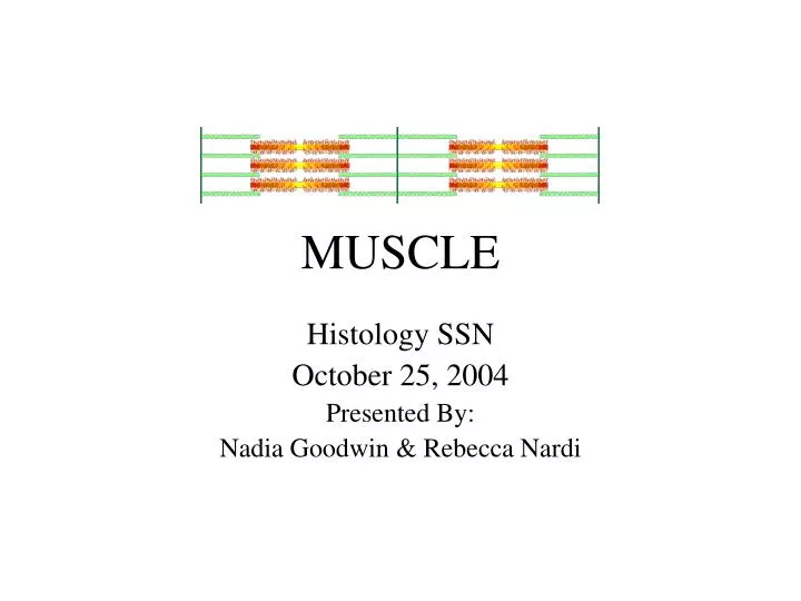
MUSCLE. Histology SSN October 25, 2004 Presented By: Nadia Goodwin & Rebecca Nardi. Muscle Types. Which is Which? Cardiac? Skeletal? Smooth?. Smooth Smooth Involuntary. Skeletal Striated Voluntary. Cardiac Striated Involuntary.

E N D
MUSCLE Histology SSN October 25, 2004 Presented By: Nadia Goodwin & Rebecca Nardi
Muscle Types Which is Which? Cardiac? Skeletal? Smooth? • Smooth • Smooth • Involuntary • Skeletal • Striated • Voluntary • Cardiac • Striated • Involuntary What other places can “skeletal” muscle be found?
Skeletal Muscle Cross section • Endomysium • Perimysium • Epimysium
Skeletal Muscle Organization Ross Fig 10-3 (p.250, 4th ed.)
How do Muscles get their Stripes? • Light band is the I band = actin only • Actin anchored at the Z disc at center of I band • Dark band is the A band = myosin + overlapping actin • Contains H band = myosin only • Myosin anchored at the M line
When Muscles Contract… • The A band does NOT change length • H and I bands shrink • Z discs are drawn closer together • Actin & myosin filaments do not actually shorten, they just slide past each other
The Triad • Triad = 1 T tubule + 2 terminal cisternae of the sarcoplasmic reticulum • There are 2 triads per sarcomere (in skeletal muscle) located at the ends of the A band (A-I junctions) • Function- Depolarization of sarcolemma (T tubule) triggers Ca2+ release from sarcoplasmic reticulum to initiate contraction
Motor End Plates Motor unit = motor nerve + muscle cells it innervates
Other Things to Know Muscle-tendon Junction
Cardiac Muscle Like skeletal muscle, cardiac muscle has: • Sarcomeres (striations) • T-tubules • Calcium regulation at level of thin filaments So what’s different? Lab 7 Slide 13
Cardiac Muscle – Defining Characteristics Branching myofibrils Large, central, ovoid nuclei • Intercalated Disks Lab 7 Slide 14
Intercalated Disks Also, T Tubules and SR arranged in a diad, not triad Lab 7 Slide 16
Which is which? Skeletal Cardiac
Which is which? Lab 7. Slide 15. Cardiac Lab 17. Slide 10. Skeletal
Smooth Muscle Located in blood vessels, ducts, and here, in the GI tract. (This is the colon.) Lab 7 Slide 17
Smooth Muscle Longitudinal section Myenteric Ganglion ? Cross Section
A Closer Look Elongated nuclei Indistinct boundaries (w/ H&E) Wrinkled nuclei Centrally located nuclei Lab 7 Slide19
Smooth Muscle Cardiac Muscle
Smooth Muscle vs. CT Smooth Muscle • Darker Staining • Nuclei within fibers • Wrinkled nuclei DIACT • Lighter Staining • Fewer nuclei, outside fibers Lab 4. Slide 16.
Smooth Muscle v. CT Smooth Muscle DRACT
Silver Stain Clear Basal Lamina Lab 7 Slide 23
A Closer Look – Why the “Dotted Lines”? Interrupted basal lamina due to GAP JUNCTIONS • Ensure coordination during contraction Lab 7 Slide 24
What Muscle?? Smooth Nerve Skeletal Esophagus – Where Smooth and Skeletal Muscle Combine Lab 17. Slide 14
1. B A
3. A B C D