Download
1 / 55
550 likes | 745 Views
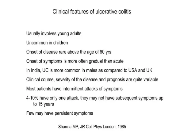
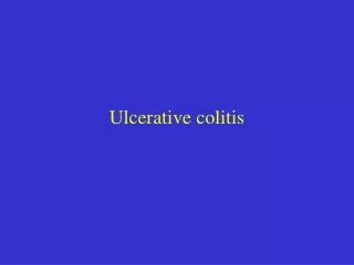
Ulcerative colitis. Disease distribution. Ulcerative Colitis. Left sided cloitis. Proctosigmoiditis. Proctitis. Disease distribution. The disease typically is most severe distally and progressively less severe more proximally.

E N D
Disease distribution Ulcerative Colitis Left sided cloitis Proctosigmoiditis Proctitis
Disease distribution • The disease typically is most severe distally and progressively less severe more proximally. • In contrast to Crohn's disease, continuous and symmetrical involvement is the hallmark of UC, with sharp transition between diseased and uninvolved segments of bowel
Physical findings • mild or even moderately severe disease: -few abnormal physical signs • severe attacks : -tachycardia -fever -orthostasis -weight loss • fulminantcolitis: - the abdomen often becomes distended and firm, with absent bowel sounds and signs of peritoneal inflammation.
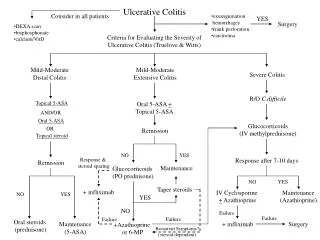
Colectomy in Ulcerative colitis • The probability of colectomy is highest in the first year of diagnosis • the overall colectomy rate is 24% at 10 years and 30% at 25 years • The probability of colectomy is related to the extent of disease at diagnosis.
Diagnosis • No single test allows the diagnosis of UC with acceptable sensitivity and specificity. • the diagnosis relies on a combination of : -compatible clinical features -endoscopic appearances -histologic findings. • Stool cultures should be obtained to exclude infectious colitis
Diagnosis • colonoscopy should be performed to establish the extent of the disease and to exclude Crohn's disease. • Multiple biopsy specimens should be taken from throughout the colon to map the histologic extent of disease and to confirm the diagnosis if there is concern about Crohn's disease. • Additionally, intubation and biopsy of the terminal ileum should be attempted to exclude the presence of Crohn's disease.
Endoscopic findings • Strictures occasionally may be present in patients with chronic UC • Caused by focal muscular hypertrophy associated with inflammation. • Malignancy must be excluded in patients with UC who have strictures, particularly those with long strictures without associated inflammation and those proximal to the splenic flexure.
Radiology: Barium enema • less frequently used in the care of patients with UC • may be superior to colonoscopy for certain indications
Assessment of disease severity • Mild <4 stools/day, without or with only small amounts of mucus No blood No fever No tachycardia Mild anemia ESR < 30 mm/hr • Moderate Intermediate between mild and severe • Severe >6 stools/day, with blood Fever > 37.5°C Heart rate > 90 beats/min Anemia with hemoglobin < 75% of normal
Mayo score • A numerical disease activity instrument • It is the sum of scores from four components • It ranges from 0 to 12, with the higher total score indicating a more severe disease
Mayo score • Remission: score <2 • severe disease: score> 10 • Clinical response: decrease by 3 points from the patient's initial baseline score.
Fulminant colitis • Patients with severe fulminant colitis: - appear toxic -fever higher than 101°F -tachycardia - abdominal distention -signs of localized or generalized peritonitis -leukocytosis • Toxic megacolon: radiologic evidence of colon dilatation to greater than 6 cm in an acutely ill patient. • Fulminant colitis and toxic megacolon are clinical diagnoses, and endoscopic examination should be avoided in patients with severe or fulminant colitis because of the risk of inducing megacolon or perforation.
Extraintestinal manifestations • numerous complications may occur distant from the bowel • Many of these complications are common to both Crohn's disease and ulcerative colitis • In large series, extraintestinal manifestations are found to occur more frequently in Crohn's disease than in ulcerative colitis and are more common among patients with colonic involvement than in patients with no colonic inflammation • one fourth of all patients with Crohn's disease will have an extraintestinal manifestation of IBD.
Musculoskeletal Manifestations • Among the most common extraintestinal manifestations are disorders of the bones and joints • In most patients, joint symptoms occurred in the setting of a relapse of bowel symptoms • Among patients with Crohn's disease, nearly one half had joint symptoms in association with a relapse in bowel disease.
Musculoskeletal Manifestations • Axial arthropathy occurs less frequently than does peripheral arthropathy in patients with IBD, and includes sacroiliitis and spondylitis. • Spondylitis associated with IBD presents as insidious low back pain and morning stiffness that is improved by exercise. • Does not parallel the activity of bowel disease
Skin: pyoderma gangrenosum • The most common skin lesions associated with IBD are pyoderma gangrenosum and erythema nodosum. • Neither condition is found solely in IBD, and the finding of one or the other lesion is not specific for either major form of IBD.
Skin: pyoderma gangrenosum • Pyodermagangrenosum appears first as a papule, pustule, or nodule and progresses to an ulcer with undermined borders. The ulcer typically has a violaceous rim and crater-like holes pitting the base • most often appears on the leg however it can occur virtually anywhere on the body. • Rare, occurs in 1-2% of patients • In Crohn's disease pyodermagangrenosum often occurs without an associated flare of bowel symptoms.
Mucocutaneous Manifestations • Aphthous ulcers of the mouth are common among patients with Crohn's disease and ulcerative colitis • These lesions usually occur with flares of colitis and resolve on control of the bowel disease • Angular cheilitis is seen in nearly 8% of patients with Crohn's disease. • Angular stomatitis and a sore tongue may be seen in patients with deficiencies of iron or other micronutrients
Ocular Manifestations:episcleritis • estimated to occur in 6% of patients with Crohn's disease, 5% of patients with ulcerative colits • consists of painless hyperemia of the sclera and conjunctiva with no affection of visual acuity. • It typically parallels the activity of bowel disease and usually responds to anti-inflammatory therapy
Ocular Manifestations: uveitis • uveitis presents as an acute or subacute painful eye with visual blurring and often photophobia and headache. Visual acuity is preserved unless the posterior segment becomes involved. • Temporal correlation of uveitis with the activity of the colitis is less predictable than with episcleritis. • Uveitis should receive prompt treatment with local steroid ocular drops to prevent progression to blindness.
Hepatobiliary Manifestations • Gallstones are found in more than 25% of men and women with Crohn's disease, representing a relative risk of 1.8 compared with the general population. • Asymptomatic and mild elevations of liver biochemical tests often are seen in IBD. In most cases, the levels return to normal once remission is achieved. These abnormalities are thought to be related to a combination of factors, including malnutrition, sepsis, and fatty liver. • Primary sclerosingcholangitismore often is associated with ulcerative colitis but may occur in 4% of patients with Crohn's disease, usually those with colonic involvement.
Hepatobiliary Manifestations :PSC • PSC should be excluded in patients with UC who have persistently abnormal liver tests or evidence of chronic liver disease. • PSC is independent of the underlying colitis and it usually follows a progressive course after many years of stable disease. • Unfortunately, no treatment has been shown definitively to be effective.