Download

1 / 36
400 likes | 1.56k Views
Complicated Corneal Ulcer. Perforated Corneal Ulcer. Healed Keratocele . Hypopyon Ulcer. TypesCorneal Ulcer (Superficial Purulent Keratitis) with Hypopyon Ulcer Serpen. Hypopyon Ulcer. There is always an associated iritis in all cases of Corneal Ulcer due to diffusion of toxins of infecting bacteria into the eye.Sometimes iridocyclitis is so severe that it is accompanied by outpouring of leucocytes from uveal blood vessels and these cells gravitate to bottom of the anterior chamber to form h30157

E N D
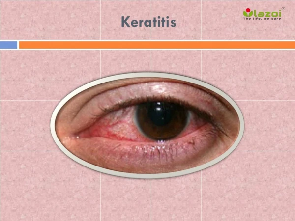
1. Keratitis
2. Complicated Corneal Ulcer
4. Healed Keratocele
5. Hypopyon Ulcer Types
Corneal Ulcer (Superficial Purulent Keratitis) with Hypopyon
Ulcer Serpen
6. Hypopyon Ulcer There is always an associated iritis in all cases of Corneal Ulcer due to diffusion of toxins of infecting bacteria into the eye.
Sometimes iridocyclitis is so severe that it is accompanied by outpouring of leucocytes from uveal blood vessels and these cells gravitate to bottom of the anterior chamber to form hypopyon (pus in anterior chamber)
7. Introduction The hypopyon which forms in bacterial keratitis is sterile as the leucocyte secretion is due to irritation by toxins and not by the bacteria
Hypopyon may develop in hours and it may change in quantity and may also rapidly disappear.
Hypopyon in bacterial keratitis is fluid and changes its position with change in head posture
8. Etiology
9. Predisposing Factors High Virulence of infecting organism
Resistance of the tissues, which is low
Dacryocystitis
Ocular trauma
Old, debilitated or alcoholic
Measles or scarlet fever
10. Organisms Pyogenic organisms like Staphylococci, Streptococci, Gonococci, Moraxella, Pseudomonas and Pneumococci
11. Hypopyon Ulcer
12. Ulcus Serpen Ulcus Serpen is hypopyon ulcer caused by Pneumococci in adults and has tendency to creep over the cornea in serpiginous fashion
13. Symptoms Sever pain, photophobia, marked diminution of vision, watering, foreign body sensation (grittiness)
14. Signs Grayish white or yellowish disc like lesion near centre of cornea. Opacity is marked at edges than at the centre and more marked in one direction (where it is progressive). In the direction of progression there is cloudiness (grey coloured) and fine line ahead of disc
Cornea may be lusterless. There is severe iritis and aqueous is hazy or there may be rank hypopyon amount which varies
15. Signs Untreated ulcer increases in depth and spread towards the side of dense infiltration, while on the other side simultaneously healing (cicatrization) takes place.
There is infiltration just anterior to Descemets� membrane underneath the floor of ulcer with normal intervening lamellae, due to which there is tendency for perforation of cornea. Intra-ocular tension is usually raised in these cases.
16. Complications Untreated cases progresses to increase in hypopyon which becomes fibrinous leading to perforation ? Iris prolapse through large opening ?whole cornea may slough leaving peripheral cornea which is nourished by limbal vascular loops. Eventually panophthalmitis develops which destroys the eye
17. Treatment Routine treatment of Corneal Ulcer
Tab Acetazolamide
Local Betablocker
Therapeutic keratoplasty
Control of infection results in absorption of hypopyon
18. Fungal Keratitis
19. Fungal Keratitis Fungal keratitis is challenging corneal disease and presents as very difficult form bacterial keratitis. Difficulty arise in making correct clinical and laboratory diagnosis. The treatment of fungal keratitis is also difficult due to poor availability of antifungal drugs and delay in starting treatment.
Treatment is required on long term basis, intensively and often cases require therapeutic keratoplasty.
20. Fungal Keratitis Fungi enter into corneal stroma through epithelial defect, which may be due to trauma, contact lens wear, bad ocular surface or previous corneal surgery.
In stroma fungi multiply and causes tissue necrosis and inflammatory reaction.
Organisms enter deep into the stroma and through an intact Descemets membrane into the anterior chamber and iris. They can also involve Sclera.
21. Fungal Keratitis The spread is due to the fact that the blood borne growth inhibiting factors may not reach the avascular tissue like cornea and sclera.
22. Risk Factors Trauma outdoor/ or the one which involves plant matter (including contact lenses)
Topical medications: corticosteroids, anaesthetic drug abuse and topical broad spectrum antibiotics use for long time (resulting in non-competitive environment for growth)
23. Risk Factors 3. Systemic use of steroids
4. Corneal surgeries (Penetrating keratoplasty, refractive surgery)
5. Chronic keratitis (herpes simplex, herpes zoster, Vernal or allergic keratoconjunctivitis, and neurotrophic ulcer)
6. Diabetes , Chronically ill / hospitalised patients, AIDS and leprosy
24. Causative fungi Yeast: Candida species (albicans), Cryptococcus
Filamentous septated
A. Non-pigmented hyphae: Fusarium species (solani), Aspergillus species (fumigatus, flavus, niger)
B. Pigmented hyphae (dematiaceous): Alternaria, Curularia , Cladosporium species
25. Causative fungi III. Filamentous non-septated : Mucor and Rhizopus species
IV. Diphasic forms: Histoplasma, Coccidiodes, Blastomyces
26. Clinical Features
27. Symptoms
Onset is slow
Symptoms are less compared to signs
Diminution of vision, pain, foreign body sensation
28. Signs Diminution of vision, depending on location of ulcer
Conjunctival and ciliary congestion
Epithelial defect
Stromal infiltrates
Elevated areas, hypate (branching) ulcers, irregular feathery margins
Dry and rough texture
29. Fungal Keratitis with Hypopyon
30. Signs Satellite lesions
Brown pigmentation due to dematiaceous fungus (Curvularia lunata)
Intact epithelium with stromal infiltrates
Anterior chamber reaction
31. Fungal Keratitis
32. Case of Fungal+ Bacterial Keratitis
33. Laboratory Diagnosis The Gram and Giemsa stains are used as initial stains
Potassium Hydroxide (10-20 %) wet mounts
Culture Media: Sheep blood agar, Chocolate agar, Sabouraud dextrose agar, Thioglycollate broth
Anterior chamber tap under aseptic conditions to aspirate hypopyon and or endothelial plaque
34. Treatment Natamycin 5% suspension: frequency will depend on severity of condition
Candida species respond better to Amphotericin B 0.15%
Fluconazole 2%
Miconazole 1%
Scrapping every 24 to 48 hours
Treatment is required for 4 � 6 weeks
35. Treatment Sub-conjunctival injection of Miconazole 5 � 10 mgm of 10 mgm/ml suspension (indicated in severe form of keratitis, scleritis and endophthalmitis)
Systemic:
Fluconazole or Ketoconazole is indicated in severe form of keratitis, scleritis and endophthalmitis
36. Surgical Treatment Daily debridement with spatula/ blade every 24 � 48 hours
Surgical treatment is required in approximately 1/3rd cases of fungal keratitis due to failure of medical treatment or perforation
Surgical treatment in the form of :
therapeutic keratoplasty, conjunctival flap or lamellar keratoplasty
37. Surgical Treatment Surgery is usually indicated within 4 weeks due to failure of medical treatment or recurrence of infection
Unfavorable outcome is due to scleritis, endophthalmitis and recurrence
Cryotherapy with topical antifungal treatment or corneoscleral graft in cases of fungal scleritis and keratoscleritis