Download
1 / 29
310 likes | 1.27k Views
Chronic Superficial Keratitis (Pannus). Pannus—superficial corneal vascularization/scar tissue Progressive, bilateral, can result in blindness Cause Thought to be immune-mediated (Infiltration of cornea with lymphocytes, plasma cells)

E N D
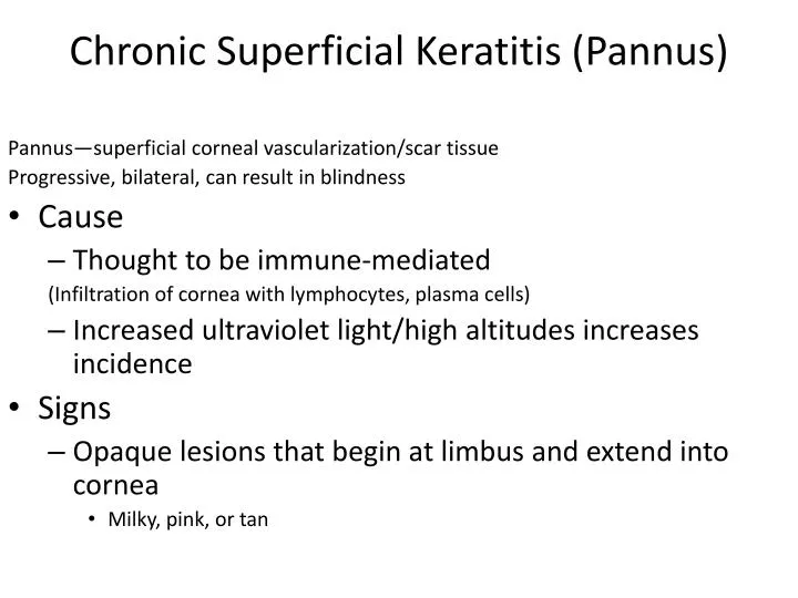
Chronic Superficial Keratitis (Pannus) Pannus—superficial corneal vascularization/scar tissue Progressive, bilateral, can result in blindness • Cause • Thought to be immune-mediated (Infiltration of cornea with lymphocytes, plasma cells) • Increased ultraviolet light/high altitudes increases incidence • Signs • Opaque lesions that begin at limbus and extend into cornea • Milky, pink, or tan
Chronic Superficial Keratitis (Pannus) • Breeds • Ger. Shep, B. Collie, greyhound, Sib. Husky • Dx • r/o KCS, corneal ulcers • Rx • Corticosteroids often lifelong • Cyclosporine often lifelong • Antibiotic eye ointment • Client info • No cure • If Rx is stopped, disease will return and progress • High altitudes and ↑sun predispose animals
Keratoconjunctivitis Sicca (KCS) Lack of tear production; tears lubricate, nourish, ↓bacteria, aid in healing Tears from 2 glands: 70%--Lacrimal gland; 30%--Nictitans gland • Signs • Recurrent conjunctivitis, corneal ulcers, keratitis • Dull, dry, irregular cornea, conjunctiva • Tenacious, mucoid ocular discharge • Blepharospasm • Crusty nares • Rx • Tear stimulation—cyclosporine, pilocarpine • Artificial tears • Client info • Px is guarded for resolution • Failure to treat → blindness
Cataracts Opacity of lens that causes reduced vision; most common disease of lens • Cause • Genetic • 2º to: • Diabetes mellitus (bilat; within 1 y of disease; ↑glucose → ↑fluid in lens) • Most common cause • Trauma (unilateral; HBC, thorn penetration, shotgun pellet) • Lens luxation • Nutritional deficiency • Uveitis • Hypocalcemia • Electrical shock • Rx • Surgical removal of lens • Treat underlying cause (e.g., Diabetes) • Client info • Most cataracts are inherited, so don’t breed affected dogs • Dogs can live quality lives even with bilat. cataracts
Cataracts • Signs • Progressive loss of vision • Opaque pupillary opening • Dx • Must be distinguished from senile nuclear sclerosis • Normal old age change; graying of lens; bilat; usually does not affect sight
Progressive Retinal Atrophy • A group of hereditary disorders causing loss of rods, cones, and/or blood supply • Breeds • Toy/min. Poodle, G. Ret, I. Set, C. Span, Schnauzer, Collie, Samoyed, N. Elkhound • Recessive gene isolated in some breeds • Signs—slow onset of blindness • Loss of night vision (rods) → loss of day vision (cones) → cataracts (±) • Dx • r/o metabolic disorders that could cause cataracts • Ophth exam • gray, granular appearance of retina • Hyperreflective retina • Vascular attenuation, optic nerve atrophy
PROGRESSIVE RETINAL ATROPHY Normal canine retina PRA, optic nerve atropy and vessel attenuation
Progressive Retinal Atrophy • Rx • None • Client info • This is an inherited disease • Avoid buying affected breeds • Have ophth exam by board certified ophth to r/o PRA • Blind animals adapt well • Have trouble in strange surroundings • Cats need well balanced diet • Taurine deficiency can lead to PRA
Anterior Uveitis • Inflammation of uvea: ciliary body, iris, choroid • Causes • Inflammation/infection – FeLV/FIP, fungal, bacterial • Neoplasia • Trauma
Uveitis – Clinical Signs • Blepharospasm • Aqueous flare – increased turbidity of aqueous humor • Miosis of affected eye • Iridal swelling or congestion • Keratic precipitates • Ciliary flush in limbal region • +/- Corneal edema • +/- hyphema
Anterior Uveitis – Treatment • Topical steroids or • Topical Anti-inflmmatory drugs (ocufen) • Or systemic steroids • Atropine – dilates eye, decreases pain • Antibiotics – topically +/- systemically
Anterior Uveitis – Client Info • Recheck within 3 days • Secondary glaucoma is frequent complication • Prognosis depends on cause • Treat for 2 months regardless of cause – blood-aqueous barrier disrupted for 6 weeks
Proptosed Globe • Cause • Trauma • Conformation • Retrobulbar abscess or neoplasia • Clinical Signs • Protrusion of the globe, • Eyelids unable to close, may be trapped behind globe
Prognosis • Favorable • brachycephalic dog, • positive direct or consensual pupillary light response • normal findings on posterior segment exam • proptosed eye with vision on initial presentation • Unfavorable indicators • non-brachycephalic • cat breed • hyphema, • no visible pupil • facial fractures • optic nerve damage and avulsion of 3 or more extraocular muscles
Proptosed Globe – Treatment • Lubricate immediately • Reduce the globe into the socket ASAP to reduce trauma to optic nerve • Enucleation if optic nerve severed • Systemic and topical antibitics • +/- Steroids
References • http://www.vetmed.ucdavis.edu/courses/vet_eyes/ • http://vanat.cvm.umn.edu/carnLabs/Lab24/Lab24.html • Alleice Summers, Common Diseases of Companion Animals • http://www.vetmed.wisc.edu/Data/CourseMaterial/Miller/Emergencies.pdf