Download
1 / 28
290 likes | 647 Views
REOVIRUS, RHABDOVIRUS & GASTROINTESTINAL VIRUSES. FE A. BARTOLOME, MD Department of Microbiology & Parasitology Our Lady of Fatima University. Reoviridae. respiratory and enteric viruses not associated with any known disease process R espiratory, E nteric, O rphan Members:

E N D
REOVIRUS, RHABDOVIRUS & GASTROINTESTINAL VIRUSES FE A. BARTOLOME, MD Department of Microbiology & Parasitology Our Lady of Fatima University
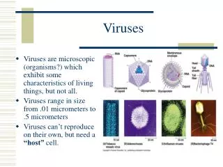
Reoviridae • respiratory and enteric viruses not associated with any known disease process Respiratory, Enteric, Orphan • Members: • Orthoreovirus – mild URT illness, GIT illness, biliary atresia • Orbivirus/Coltivirus – febrile illness associated with headache and myalgia (zoonosis) • Rotavirus – GIT illness, respiratory tract illness (?) • Non-enveloped; double-layered protein capsids with dsRNA genomes (“double:double”) • Stable over wide pH & temperature changes & in airborne aerosols
Reoviridae • STRUCTURE: • Icosahedral with double stranded segmented genome • Reovirus – 10 segments • Rotavirus – 11 segments • (+) re-assortment of gene segments create hybrid viruses • Outer capsid composed of structural proteins • Genomic segments encode structural and non-structural proteins
Reoviridae REPLICATION: Ingestion Proteolytic cleavage of outer capsid in GIT Formation of intermediate/infectious viral particle (ISVP) ISVP release core into cytoplasm Enzymes in core initiate mRNA production using + strand as template
Reoviridae • REPLICATION: • Occurs in the cytoplasm • dsRNA remains in the core • Inner capsid with complete transcription system, including enzymes for 5’ capping and polyadenylate addition • Virus leaves the cell during cell lysis
Orthoreovirus • Mammalian reovirus • Ubiquitous present in sewage & river water • 3 serotypes (1, 2 & 3) based on neutralization and hemagglutination-inhibition tests • Most people infected during childhood (+) antibodies in 75% of adults
Orthoreovirus PATHOGENESIS & IMMUNITY • No significant disease in humans • After ingestion & proteolytic production of ISVP, binds to M cells in small intestines transfer virus to lymphoid tissue of Peyer’s patches replicate (+) viremia • (+) humoral & cellular immune response to outer capsid protein
CLINICAL SYNDROMES: • Usually asymptomatic • Common cold-like mild upper respiratory tract illness • Gastrointestinal disease • Biliary atresia LABORATORY DIAGNOSIS: • Assay of viral antigen or RNA in clinical material • Virus isolation • Serologic assays Orthoreovirus
Rotavirus • Rota “wheel” • One of the most common agents of infantile diarrhea worldwide • Ubiquitous worldwide • 95% of children infected by 3 – 5 years old • Stable at: room temperature, treatment with detergents, pH 3.5 – 10, repeated freezing & thawing; survives on fomites • Divided into: • Serotypes – based primarily on VP7 outer capsid protein • Groups – based on antigenicity of VP6 & electrophoretic mobility of genomic segments A to G group A causes human disease
Rotavirus • PATHOGENESIS: • MOT: fecal-oral, possibly respiratory route • Adsorption to columnar epithelial cells covering villi of SI release of NSP4 protein (+) cytolytic & toxin-like activity loss of electrolytes & prevention of water re-absorption watery diarrhea severe dehydration
Rotavirus • NSP4 protein promotes: • calcium influx into enterocytes • release of neuronal activators • neuronal alteration in water absorption • Shortening and blunting of microvilli; mononuclear cell infiltration into lamina propia • 1010 viral particles/gm of stool released during disease maximal shedding 2 – 5 days after start of diarrhea • (+) outbreaks in pre-schools and daycare centers
Rotavirus • CLINICAL SYNDROMES: • I.P. = 48 hrs • self-limited • vomiting, diarrhea, fever, dehydration • (-) fecal leukocytes and blood • may be fatal in infants from developing countries & who are malnourished and dehydrated before the infection
Rotavirus • LABORATORY DIAGNOSIS: • direct detection of viral antigens in stool – method of choice • enzyme immunoassay • latex agglutination • serology – four fold increase in antibody titer
Coltivirus & Orbivirus • features different from other Reoviridae: • Orbivirus – outer capsid without discernible capsomeric structure; inner capsid icosahedral • Causes viremia – long lasting • Infects erythrocyte precursors without damaging them remains within the cells protected from immune response (+) viremia
Coltivirus & Orbivirus • Coltivirus: • causes Colorado Tick Fever • vector: Dermacentor andersoni (wood tick) • one of the most common tick-borne diseases in the U.S. • symptoms of acute disease resemble dengue • infect vascular endothelial & vascular smooth muscle cells & pericytes weak capillary structure (+) hemorrhage hypotension shock • neuronal infection – meningitis & encephalitis • (+) leukopenia involving both neutrophils and lymphocytes HALLMARK
Coltivirus & Orbivirus • LABORATORY: • Immunofluorescence – most rapid and best technique detection of viral antigen on surface of red blood cells on blood smear • Antibody titer – specific IgM (+) 45 days after onset of illness presumptive evidence of acute or very recent infection
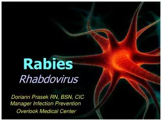
Rhabdovirus • “rhabdo” rod • bullet-shaped; enveloped; icosahedral nucleocapsid • negative ssRNA prototype for replication of negative stranded enveloped viruses
Rhabdovirus • encode 5 proteins: • G – synthesized by membrane-bound ribosomes; attach to host cell & internalized by endocytosis; generates neutralizing antibodies; prototype for studying eukaryotic glycoprotein processing • N – major structural protein; protects the RNA from ribonuclease digestion • L and NS – constitute the RNA-dependent RNA pol • M – matrix protein; lies between envelope & nucleocapsid
Rhabdovirus • RABIES VIRUS • most significant pathogen • reservoir: wild animals • source of virus: • Major – saliva in bite of rabid animal • Minor – aerosols in bat caves containing rabid bats • found worldwide; no seasonal incidence
Rhabdovirus • PATHOGENESIS & IMMUNITY • MOT: 1. bite of rabid animal • 2. inhalation of aerosolized virus • 3. transplanted infected tissue (e.g. cornea) • 4. inoculation through intact mucosal membrane • rabies infection of animal cause secretion of the virus in the animal’s saliva & promotes aggressive behavior “mad dog” promote transmission • not very cytolytic remains cell associated
Wks to mos Travel by retrograde axoplasmic transport to DRG & to SC Prodrome phase Afferent neurons CNS (hippocampus, brainstem, ganglionic cells of pontine nuclei, Purkinje cells of cerebellum) Rhabdovirus MOT Muscle at site of inoculation Bind to nicotinic Ach or ganglioside receptors of neurons Replicate at site of bite Incubation phase Skin of head & neck, salivary glands, retina, cornea, nasal mucosa, adrenal medulla, renal parenchyma, pancreatic acinar cells Neurologic phase
Rhabdovirus • length of incubation period determined by: • concentration of virus in inoculum • proximity of wound to brain • severity of the wound • host’s age • host’s immune status • rarely causes inflammatory lesions • CMI with little or no role in protection • antibody can block spread of virus to CNS if administered or generated during the incubation period • long incubation period allows active immunization as post- exposure treatment
Rhabdovirus PROGRESSION OF RABIES DISEASE
Rhabdovirus • LABORATORY DIAGNOSIS: • done to confirm the diagnosis • 1. detection of viral antigen in CNS or skin via immunofluorescence most widely used • 2. virus isolation – cell culture • 3. serology – antibody titers in serum and CSF • 4. detection of Negri bodies – intracytoplasmic inclusions containing aggregates of viral nucleocapsids in affected neurons HALLMARK
Rhabdovirus • PRE-EXPOSURE VACCINATION: • HDCV IM or intradermally x 3 doses 2 years protection • POST-EXPOSURE PROPHYLAXIS: • local treatment of wound – wash immediately with soap & water • WHO Expert Committee on Rabies – instillation of anti-rabies around the wound • active + passive immunization • HDCV IM on day of exposure then on days 3, 7, 14 & 28 + 1 dose human rabies immune globulin (HRIG)
Gastrointestinal Viruses CALICIVIRUSES • 5 groups: 1. Norwalk 4. Marine virus 2. Sapporo viruses 5. Rabbit hemorrhagic dse virus 3. Hepatitis E • approx. same size as Picornaviruses • naked, positive sense ssRNA viruses • viruses distinguishable by capsid morphology • compromise function of intestinal brush border prevent re-absorption of water and nutrients • cause outbreaks of gastroenteritis
Gastrointestinal Viruses • I.P. = 24 – 48 hrs • MOT: 1. fecal-oral – water, shellfish, food service 2. possible airborne • immunity short-lived & not protective • symptoms similar to Rotavirus infection - Norwalk & related viruses diarrhea, n & v, esp. in children; fever in 1/3 of patients • resolve within 12 – 60 hrs
Gastrointestinal Viruses ASTROVIRUSES • seen in stools from infants & young children with diarrhea • may be shed in extraordinarily large amounts in feces • associated with diarrhea in young children in daycare centers • symptoms similar to Norwalk but without vomiting • minimally pathogenic ADENOVIRUS • replicates in intestinal cells • adenovirus group F serotypes 40 & 41 infantile gastroenteritis detected by electron microscopy or antigen-based assays • usually sub-clinical