Download
1 / 74
1.01k likes | 1.74k Views
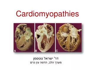
Reversible Cardiomyopathies. Dr Sajeer K T Senior Resident, Dept. of Cardiology, MCH Calicut. Dilated cardiomyopathy (DCM) : - characterized by dilation and impaired contraction of one or both ventricles. DCM - caused by a variety of specific diseases.

E N D
Reversible Cardiomyopathies Dr Sajeer K T Senior Resident, Dept. of Cardiology, MCH Calicut
Dilated cardiomyopathy (DCM) : - characterized by dilation and impaired contraction of one or both ventricles DCM - caused by a variety of specific diseases ≥ 50% of patients with DCM: - an etiologic basis will not be identified - Idiopathic DCM • One series- 1278 patients with congestive heart failure • Idiopathic — 51 percent • Idiopathic myocarditis — 9 percent • Occult coronary disease — 8 percent • Other identifiable causes — 32 percent Felker, et al. The spectrum of dilated cardiomyopathy. The Johns Hopkins experience with 1,278 patients. Medicine (Baltimore) 1999; 78:270
Clinical Presentation - Most patients present between age : 20-60 years - Can occur in children and elderly - Symptoms of CHF - Incidental detection of asymptomatic cardiomegaly - Symptoms related to coexisting arrhythmia, conduction disturbance - Thromboembolic complications - Sudden death
Reversible causes of dilated cardiomyopathy • Peripartum cardiomyopathy • Tachycardia-mediated cardiomyopathy • Takotsubo cardiomyopathy • Alcoholic cardiomyopathy • Cocaine • Medications • Ischemia • Endocrine dysfunction • SLE, Sarcoidosis • Nutritional deficiencies • Electrolyte abnormalities • Obstructive sleep apnea
Peripartum cardiomyopathy (PPCM) • Cardiomyopathy manifesting between the last month of pregnancy • and 6 months postpartum - Toxic postpartal heart failure, Meadows synd., Zaria synd. - Incidence in published series of 1:1300 to 1:4000 live births Criteria for PPCM : (1) Development of CHF secondary to deceased LV systolic function in the last month of pregnancy or within 5 months after delivery (2) Absence of pre-exciting cardiac dysfunction (3) Absence of determinable cause of cardiomyopathy (4) LV systolic dysfunction: - EF < 45% - Fractional shortening of less than 30% - LV end diastolic dimension of greater than 2.7 cm/m2
- Risk factors for PPCM: • Advanced age (>30 years) • Multiparity • Twin gestation • Use of tocolytic therapy • Preeclampsia • Obesity • Chronic hypertension • Black race - Etiology - currently unknown - Current hypotheses are: (1) infectious (2) immunologic (3) nutritional (4) drug-induced (5) familial
Infectious: Felker et al : demonstrated a 62% prevalence of myocarditis in patients PPCM who underwent endomyocardial biopsy Immunologic: - significant elevations of TNF-Alpha, IL-6 and Fas/APO-1 in patients with PPCM - antibodies formed against actin can cross react with myocardium - antibodies to cardiac muscle proteins have been identified in patients with PPCM Nutritional: - Low selenium levels - a possible risk factor for PPCM Drug-induced: - prolonged terbutaline therapy Familial:
Echocardiography remains the tool for evaluation and follow-up for women with postpartum cardiomyopathy - Myocardial LV dysfunction - decrease in LV EF or FS - LV thrombus is common with a LVEF less than 35% Normal systolic function excludes postpartum cardiomyopathy - should lead to an evaluation for high output failure - anaemia - thyrotoxicosis
Treatment Initial treatment of PPCM is similar to treatment for other forms of heart failure - diuretics, vasodilators (hydralazine and nitrates), digoxin - (ACEI contraindicated in pregnancy) • Immunosuppression: • Pentoxifylline: inhibit proinflammatory cytokines • (TNF-alpha, CRP, and Fas/Apo-133 • - Intravenous immune globulin Immunosuppressive therapy : - considered if an endomyocardial biopsy indicates the presence of myocarditis - if there is no improvement after 2 weeks of standard HF therapy
Outcome and prognosis - Reported mortality rate for PPCM ranges from 15% to 50% - Cardiac transplantation is an option if the patient fails to improve - no consensus on whether subsequent pregnancies should be avoided by women who survive PPCM
Takotsubo Cardiomyopathy A New Form of Acute, Reversible Heart Failure
a condition featuring symptoms and signs of AMI without • demonstrable coronary artery stenosis or spasm in which the • heart takes on the appearance of a Japanese octopus fishing pot • called a “takotsubo” • Left ventricular dysfunction can be remarkably • depressed, recovers within a few weeks - occurs predominantly in postmenopausal women soon after exposure to sudden unexpected emotional or physical stress. Sato et al (1990) first described - reversible cardiomyopathy - tako-tsubo-like left ventricular dysfunction - Apical ballooning or stress cardiomyopathy
- noncardiac surgery is one of the most frequent trigger for cardiovascular events ( highest risk in those undergoing vascular surgery due to coexisting severe coronary artery disease) - incidence of ABS : 1% to 2% of patients with an acute MI
Diagnostic Criteria Mayo Clinic proposed diagnostic criteria in 2004 (1) - transient hypokinesis, akinesis, or dyskinesis in the LV mid segments with or without apical involvement - regional wall motion abnormalities that extend beyond a single epicardial vascular distribution - a stressful trigger (2) - the absence of obstructive coronary disease or angiographic evidence of acute plaque rupture (3) - new ECG abnormalities (ST-segment elevation and/or T-wave inversion) or modest elevation in cardiac troponin (4) - absence of pheochromocytoma and myocarditis
syndrome much more common in women • typically preceded by exposure to emotional or physical stressors • - most frequent clinical symptoms on admission are chest pain and • dyspnea resembling AMI • - most common ECG findings on admission : ST elevation in • precordial leads • high levels of serum catecholamines and of plasma BNP • Cardiac enzyme levels (eg, CKMB , Tn T) - slightly increased. - Cardiac magnetic resonance imaging: -lack of delayed contrast enhancement
RV may develop similar regional wall motion abnormality in 30% of patients - develop congestive heart failure Inverted Tako-tsubo : - a rare variant presents with hypokinesis of the base of the heart with preserved apical function
- Cine sequences of CMR imaging • during systole (A) and diastole (B) • in the acute phase. • Normal function could be documented • after 3 weeks (C, systole; D, diastole) • Contrast-enhanced CMR image did not • show myocardial hyper enhancement • even in the delayed phase (E).
Pathophysiology: Myocardial biopsy: - interstitial infiltrates( mononuclear lymphocytes, leukocytes, and macrophages) - myocardial fibrosis - contraction bands with or without overt myocyte necrosis Coagulation necrosis : seen in myocardial infarction • Catecholamine Cardio toxicity: • - high local concentrations of nor-epinephrine might evoke • basal hyperkinesis • - increasing mechanical wall stress at the apex • - increasing end-diastolic pressure and BNP levels
A Pathogenetic Concept About Takotsubo Cardiomyopathy Possible underlying mechanism of classic takotsubo cardiomyopathy
Prognosis • Prognosis is generally favourable • Fatal complication • LV free wall rupture • heart failure, with or without pulmonary oedema - Recurrence rate of takotsubo cardiomyopathy : 10% - Complete recovery - by 4 to 8 weeks - Inhospital mortality from ABS is very low (1- 2%)
Tachycardia-mediated cardiomyopathy • impairment in LV function secondary to chronic tachycardia, which • is partially or completely reversible after normalization of heart rate • and/or rhythm irregularity • ---Gallagher JJ Fenelon et al : Tachycardiomyopathy: a) Pure type b) impure Type • Pure type: • chronic tachycardia causes LV dysfunction in a normal heart and completely recovers after termination of the tachycardia • Impure Type: • occurs in patients with structural heart diseases, and cardiac dysfunction may only recover incompletely after termination of the tachycardia
Possible mechanisms for myocardial dysfunction - Myocardial energy depletion - Impaired energy utilization - Myocardial ischemia Abnormal calcium handling: - the severity of calcium cycling abnormalities correlates with the degree of ventricular dysfunction (extensive abnormalities in calcium channel activity and sarcoplasmic reticulum calcium transport ) • calcium availability to myocytes decreased • > reduction in contractility
- Tachycardiomyopathy can occur at any age - reported in infants, children ,adolescents and adults - incidence of tachycardia-induced cardiomyopathy - unknown - risk factors: - type, rate and duration of tachyarrhythmia - patient’s age - underlying heart disease - drugs - coexisting medical conditions - diagnosis of tachycardiomyopathy considered - patient with LV systolic dysfunction and chronic or frequently recurring cardiac arrhythmia
Tachycardiomyopathy is induced by various supraventricular and ventricular arrhythmias • - Ectopic atrial tachycardia (EAT) - in children • Permanent form of junctional reciprocating tachycardia (PJRT) • - often incessant and refractory to antiarrhythmic drugs • - commonly results in tachycardiomyopathy • Fenelon et al proposed the following criteria: • 1) Dilatation of the heart or heart failure • 2) Chronic or very frequent cardiac arrhythmias, including • incessant SVTs, AF or AFL and incessant VT If chronic tachycardia continued more than 10-15% of the day, with an atrial rate of more than 150% of that predicted for age, - tachycardiomyopathy occurs.
Treatment - Basic concept for the treatment of tachycardiomyopathy - controling the heart rate - SVT : digitalis or verapamil : common drugs of choice - Class I anti-arrhythmic drugs: negative inotropic effect - clinical use should be careful - In pediatric cases, EAT or PJRT : incessant - Amiodarone - class III - dofetilide, sotalol, - ibutilide or azimilide - Catheter ablation : performed as the first-line therapy
Alcoholic cardiomyopathy • ethanol may induce asymptomatic LV systolic dysfunction • 30% of asymptomatic chronic alcoholics have evidence of LV • systolic dysfunction • - ethanol abuse is the leading cause of nonischemic DCM in • industrialized countries
- likelihood of developing an ethanol-induced DCM correlates with the amount of ethanol that is consumed in a lifetime - Men – consumption more than 80 g of ethanol/day for at least 5 yrs (1 L of wine, 8 std-sized beers, or 0.5 pint of hard liquor) -Women - more susceptible to ethanol's cardiotoxic effects than men • complete abstinence or reduction in ethanol consumption (less • than 60 g of ethanol/day): substantial improvement in LV • systolic function and symptoms of HF
Cocaine and heart Cocaine - commonest illicit drug used and the most frequent cause of drug related deaths • Cocaine related complications : • Cardiac: myocardial ischaemia, coronary artery spasm, • acute MI, myocarditis, cardiomyopathy, arrhythmia, • hypertension • Vascular: aortic dissection and rupture, vasculitis • Gastrointestinal: mesenteric ischaemia or perforation • 4. Pulmonary: pulmonary oedema, pulmonary infarction
Dilated cardiomyopathy in cocaine users: - attributable to the direct toxic effects • - heart failure results from: • - myofibrils destruction • - interstitial fibrosis • - myocardial dilatation • cocaine induced hyper adrenergic state may contribute to the • cardiomyopathy (similar to pheochromocytoma) - myocardial dysfunction is reversible with abstinence of cocaine
Medications - anthracycline-induced cardiomyopathy - most extensively studied - cardiomyopathy and clinical heart failure are the main dose limiting side effects of anthracyclines • cardiomyopathy can occur within the 1st year and up to a decade • after completion of therapy risk factors: 1 . cumulative dose of anthracyclines administered (not to exceed 450 mg/m2 in adults) 2. extremes of age 3. concomitant chemotherapy and radiation 4. history of cardiovascular disease
Endocrine dysfunction • thyroid dysfunction • excess sympathetic activity in pheochromocytoma • rarely Cushing's syndrome • growth hormone excess or deficiency • cardiac dysfunction which can usually be reversed by • correction of the endocrine disorder - hyperthyroidism or hypothyroidism: - exact mechanisms of dilated cardiomyopathy not known - thyroid hormone is known to alter preload, after load, heart rate, and contractility - excess T3 causes myocyte hypertrophy
Pheochromocytoma : - excess sympathomimetic amines cause focal direct myocyte injury - inflammation, down regulation of beta receptors - reduction of viable myofibrils Catecholamines – - increase both intracellular calcium and free radicals - contribute to myocyte injury
Ischemia - cardiomyopathy • coronary atherosclerosis is MC cause of DCM in US • 50 to 75 percent of patients with HF • usually irreversible due to myocardial infarction and subsequent • ventricular remodeling • patients with ischemic DCM with hibernating myocardium • - benefit of revascularisation • Revascularization of hibernating myocardium beneficial in • patients with ischemic DCM who have less severe heart disease
Selenium deficiency and cardiomyopathy Selenium deficiency: - identified as a factor causing HF syndromes in areas of very low selenium intakes( China ) - endemic selenium-responsive cardiomyopathy Keshan disease Selenium deficiency : - decreases the activity of glutathione peroxide - resulting in increased free radicals > toxic to cardiac myocytes Similar cases : - HIV-infected patients - subjects on parenteral nutrition - Crohn’s disease Possible causes of Keshan disease are: - viral infection - nutritional factors (insufficient Zn or molybdenum, excessive barium or lead)
Thiamine: • - plays an important role in normal oxidative phosphorylation and • myocardial energy production • - deficiency initially presents as a high output state secondary to • vasodilation • followed by eventual depression of myocardial function – • development of a low output state
Obstructive sleep apnea - cardiomyopathy • obstructive sleep apnea: contribute to the impairment of left • ventricular dysfunction • history of snoring, daytime somnolence, and obesity should alert • the clinician to the diagnosis • therapy : nasal continuous positive airway pressure during sleep • lead to a significant improvement in LV dysfunction
Electrolyte abnormalities: Chronic hypophosphatemia and hypocalcaemia: - reversible cardiomyopathy Infections : - Bacterial, spirochetal, rickettsial, fungal, protozoal, helminthic infections - implicated as causes of necrotic or inflammatory lesions - may lead to dilated cardiomyopathy