Download
1 / 73
740 likes | 1.14k Views
Digestive system. By Sindhu Priya E S. Objectives. At the end of this chapter, you will be able to: List and describe the functional anatomy of the organs and accessory organs of the digestive system

E N D
Digestive system By SindhuPriya E S
Objectives At the end of this chapter, you will be able to: • List and describe the functional anatomy of the organs and accessory organs of the digestive system • Discuss the processes and control of ingestion, propulsion, mechanical digestion, chemical digestion, absorption, and defecation • Discuss the roles of the liver, pancreas, and gallbladder in digestion • Compare and contrast the digestion of the three macronutrients
introduction Digestive system • The digestive system is the collective name used to describe the alimentary canal, some accessory organs and a variety of digestive processes which take place at different levels in the canal to prepare food eaten in the diet for absorption.
digestive processes • Ingestion • Deglutition • Propulsion : Peristalsis • Digestion • Mechanical breakdown by mastication • Chemical digestion by enzymes • Absorption • Elimination
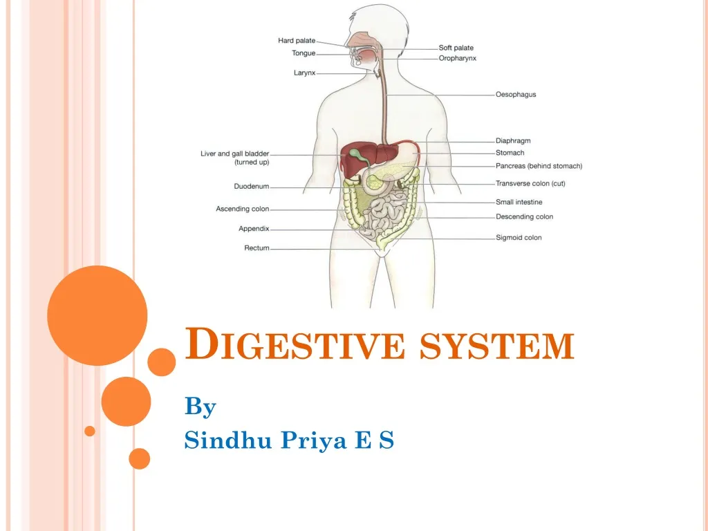
Organs of the digestive system • Mouth • Pharynx • Oesophagus • Stomach • Small intestine • Large intestine • Rectum and anal canal
Accessory organs • 3 pairs of salivary glands • Pancreas • Liver and the biliary tract BASIC STRUCTURE OF ALIMENTARY CANAL • Adventitia or outer covering: fibrous tissue or peritonium • Muscle layer: longitudinal and circular, plexus • Submucosal layer: loose connective tissue • Mucosa — lining
Secretions from glands • Saliva from the salivary glands • Gastric juice from the gastric glands • Intestinal juice from the intestinal glands • Pancreatic juice from the pancreas • Bile from the liver
Mouth Boundaries of mouth • Anteriorly —by the lips • Posteriorly —it is continuous with the oropharynx • Laterally —by the muscles of the cheeks • Superiorly —by the bony hard palate and muscular soft palate • Inferiorly —by the muscular tongue and the soft tissues of the floor of the mouth.
Tongue • External carotid artery supplies blood to tongue • Hypoglossal nerves - muscles • Lingual branch of the mandibular nerves – pain, temp & touch • Facial and glossophanryngeal nerves - taste Functions The tongue plays an important part in: • Mastication (chewing) • Deglutition (swallowing) • Speech • Taste
teeth • The teeth, or dentes (singular = dens), are organs similar to bones that you use to tear, grind, and otherwise mechanically break down food. Types of Teeth sets or dentitions • Deciduous teeth - 20 • Permanent teeth – 32 (wisdom teeth)
Salivary glands • Salivary glands are housed within the mucous membranes of the mouth and tongue • They constantly secretes 1-1.5 L of saliva per day which helps in moistening of mouth, teeth, food and aids in chemical digestion of carbohydrates • Three pairs of Salivary glands • Parotid glands • Submandibular glands • Sublingual glands
Position of the salivary glands Mumps: Enlargement and inflammation of parotid glands by paramixovirus
Saliva Composition • water • mineral salts • enzyme: salivary amylase • mucus • lysozyme • immunoglobulins • blood-clotting factors Functions • Chemical digestion of polysaccharides at 6.8 pH (pH of saliva 5.8 to 7.4) • Lubrication of food • Cleansing and lubricating • Non-specific defence • Taste Regulation of Secretion
Pharynx Parts of pharynx • Nasopharynx- respiration • Oropharynx – respiration and digestion • Laryngopharynx - respiration and digestion Wall of pharynx contains • Inner mucosal layer • Middle fibrous tissue • Outer involuntary muscular layer – swallowing Function • Helps in swallowing of bolus
oesophagus • The oesophagus is a muscular tube that connects the pharynx to the stomach. • 25.4 cm (10 in) in length, located posterior to the trachea normally collapsed when not swallowing • cricopharyngeal sphincter and cardiac or lower oesophageal sphincter are present at the ends (GERD) Structure • Outer adventitia - loose connective tissue • Middle muscle layer – skeletal and smooth muscle • Inner mucosal layer – stratified and columnar
Deglutition • Swallowing: The movement of food from the mouth to the stomach • 4-8 sec for solid foods. 1 sec for liquids • Complex process involving muscles of tongue, pharynx and oesophagus • Three stages of swallowing • Voluntary phase • Pharyngeal phase • Oesophagealphase Check the video of phases of swallowing in the following link https://www.youtube.com/watch?v=pNcV6yAfq-g
stomach Organs associated with the stomach • Anteriorly —left lobe of liver and anterior abdominal wall • Posteriorly —abdominal aorta, pancreas, spleen, left kidney and adrenal gland • Superiorly — diaphragm, oesophagus and left lobe of liver • Inferiorly — transverse colon and small intestine • To the left — diaphragm and spleen • To the right—liver and duodenum
Structure of the stomach Parts of the stomach • Cardia • Fundus • Body • Pyrolus Curvatures • Greater curvature • Lesser curvature
Types of cells in gastric glands • Parietal cells: located in middle region of the glands. Secretes HCl and intrinsic factor • Chief cells: Located primarily in the basal regions of gastric glands. It secretes pepsinogen, the inactive proenzyme form of pepsin. • Mucous neck cells: They are present in the upper part of the glands. Secretes thin, acidic mucus • Enteroendocrine cells: They secretes various harmones such as gastrin.
Gastric juice • About 2 litres of gastric juice is secreted daily Composition of gastric juice • Water • Mineral salts • Mucus secreted by goblet cells in the glands and on the stomach surface • Hydrochloric acid • Intrinsic factor • Inactive enzyme precursors: pepsinogens
Functions of gastric juice • Water :Further liquefies the food swallowed. • Hydrochloric acid: • acidifies the food and stops the action of salivary amylase • kills ingested microbes • provides the acid environment needed for effective digestion by pepsins. • Pepsinogensare activated to pepsins by hydrochloric acid and by pepsins already present in the stomach. They begin the digestion of proteins, breaking them into smaller molecules. Pepsins act most effectively at pH 1.5 to 3.5. • Intrinsic factor (a protein) is necessary for the absorption of vitamin B12 from the ileum. • Mucus prevents mechanical injury to the stomach wall by lubricating the contents. It prevents chemical injury by acting as a barrier between the stomach wall and the corrosive gastric juice. Hydrochloric acid is present in potentially damaging concentrations and pepsins digest protein.
Secretion of gastric juice Three phases of secretion of gastric juice • Cephalic phase • Gastric phase • Intestinal phase
acid production and its regulation A note on Gastric emptying
Functions of stomach • Chemical digestion — pepsins convert proteins to polypeptides • Mechanical breakdown — the three smooth muscle layers enable the stomach to act as a churn, gastric juice is added and the contents are liquefied to chyme • Non-specific defenceagainst microbes. Vomiting may be a response to ingestion of gastric irritants, e.g. microbes or chemicals • Temporary storage of food • Preparation of iron for absorption further along the tract — the acid environment of the stomach solubilises iron salts, which is required before iron can be absorbed • Production of intrinsic factor needed for absorption of vitamin B12 in the terminal ileum
Small intestine • Duodenum: 25 cm long • Hepatopancreaticampulla • Major duodenal papilla • Hepatopancreatic sphincter • Jejunum: 0.9 m • Ileum: 1.8m
Structure of small intestine 4 layers • Peritonium - Mesentry • Muscular layer • Submucosa • Mucosa • Circular folds, villi, microvilli- Brush border • Enterocytes, goblet cells • Lacteales Lymphnodes • Solitary lymphatic follicles • Aggregated lymphatic follicles (Peyer’s patch) – Intestinal malt Intestinal glands • Present in deep cervix of villi • They produce intestinal juice which is slightly alkaline (pH7.4=7.8) • 0.95-1.9 L per day
Intestinal glands It contains different cells • Enterocytes- digestion and absorption of nutrients in chyme • Goblet cells - secretion of mucus • G cells – secretes intestinal gastrin • I cells – secretes cholecystokinin which stimulates the release of pancreatic juices and bile • S cells – secretes secretin • M cells – secretes motilin which accelerates gastric emptying, stimulates intestinal peristalsis, and stimulates the production of pepsin
Composition of Intestinal juice • Water • Mucus • Mineral salts • Enzymes: enterokinase (enteropeptidases), peptidases, lipase, sucrase, maltase and lactase • Harmones : gastrin, secretin, CCK, motilin
Mechanical Digestion in the Small Intestine Segmentation
Chemical digestion in the smallintestine • When acid chyme passes into the small intestine it is mixed with pancreatic juice, bile and intestinal juice • Carbohydrates are broken down to monosaccharides • Proteins are broken down to amino acids • Fats are broken down to fatty acids and glycerol.
Pancreatic juice Composition Functions • Water • Mineral salts • Enzymes: — Amylase — Lipase • Inactive enzyme precursors: — Trypsinogen — Chymotrypsinogen — Procarboxypeptidase. Digestion of • Proteins by trypsin and chymotrypsin • Carbohydrates by amylase • Fats by lipase Pancreatic secretion is regulated by CCK
Bile Composition Functions • Bile has a pH of 8 and between 500 and 1000 ml secreted daily. It consists of: • Water • Mineral salts • Mucus • Bile salts • Bile pigments, mainly bilirubin • Cholesterol. • sodium taurocholate and sodium glycocholate, emulsify fats in the small intestine • Bile salts make fatty acids soluble, enabling both these and fat-soluble vitamins (e.g. vitamin K) to be readily absorbed. • Bilirubin excretion • Stercobilincolours and deodorises the faeces Biliary secretion is regulated by CCK
Absorption of nutrients It occurs by two processes • Diffusion.Monosaccharides, amino acids, fatty acids and glycerol diffuse slowly down their concentration gradients into the enterocytes from the intestinal lumen. • Active transport. Monosaccharides, amino acids, fatty acids and glycerol may be actively transported into the villi; this is faster than diffusion. Disaccharides, dipeptides and tripeptides are also actively transported into the enterocytes where their digestion is completed before transfer into the capillaries of the villi.
Absorption Through capillaries Through lacteal • Monosaccharides • Aminoacids • Mineral salts • Water soluble vitamins • Water • Fattyacids • Glycerol • Fat soluble vitamins
Functions of the small intestine • Onward movement of its contents which is produced by peristalsis • Secretion of intestinal juice • Completion of chemical digestion of carbohydrates, protein and fats in the enterocytes of the villi • Protection against infection by microbes that have survived the antimicrobial action of the hydrochloric acid in the stomach, by the solitary lymph follicles and aggregated lymph follicles • Secretion of the hormones cholecystokinin (CCK) and secretin • Absorption of nutrients
Large intestine • 1.5 m long Parts • Caecum: Vermiform appendix, ileocaecal valve • Colon • Ascending colon: Hepatic flexure • Transverse colon: Splenic flexure • Descending colon • Sigmoid colon: S shaped • Rectum: rectal valves • Anal canal • Internal sphincter-smooth muscles & involuntary • External sphincter-skeletal muscles& voluntary
Structure • Peritonium • Muscular layer • Longitudinal muscles • Do not form smooth and continuous layer • Collected into 3 bands called Taeniea coli at regular intervals of the colon • Circular muscles • Anal sphincters • Submucosa • Mucosal layer • Enterocytes & goblet cells • Stratified squamous epithelium in anus • lymph nodes