Download
1 / 72
840 likes | 1.12k Views
Chemistry 2. Lecture 11 Electronic spec of polyatomic molecules: chromophores and fluorescence. Assumed knowledge. Learning outcomes.

E N D
Chemistry 2 Lecture 11 Electronic spec of polyatomic molecules: chromophores and fluorescence
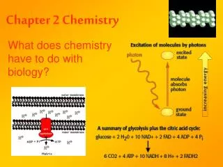
Assumed knowledge Learning outcomes • Excitations in the visible and ultraviolet correspond to excitations of electrons between orbitals. There are an infinite number of different electronic states of atoms and molecules. • Be able to draw the potential energy curves for excited electronic states in diatomics that are bound and unbound • Be able to explain the vibrational fine structure on the bands in electronic spectroscopy for bound excited states in terms of the classical Franck-Condon model • Be able to explain the appearance of the band in electronic spectroscopy for unbound excited states
Some random images of last lecture… De’ we’ De” we” re” re’ Franck-Condon Principle
Franck-Condon Principle (reprise) 6 5 4 3 2 1 0
Electronic spectra of larger molecules… A diatomic (or other small) molecule An atom A large molecule A molecule has 3N-6 different vibrational modes. When you have no selection rules any more on vibrational transitions then the spectrum quickly becomes so complicated that the vibrational states cannot be readily resolved.
Electronic spectra of larger molecules… 3s 2p 2s 1s A diatomic (or other small) molecule An atom A large molecule
Jablonski Diagrams… Vibrational levels (thin lines) Electronic states (thick lines) Etot = Eelec + Evib Again, this is the Born-Oppenheimer approximation.
Correlation between diatomic PES and Jablonski diagram Jablonski
Nomenclature and spin states In polyatomic molecules, the total electron spin, S, is one of the few good quantum numbers. If the total electron spin is zero:S = 0, then there is only one way to arrange the spins, and we have a singlet state, denoted, S (c.f. L=0 for atoms) If there is one unpaired electron: the total spin is S = ½ and there are 2 ways the spin can be aligned (up and down), and we have a doublet state, denoted, D If there are two unpaired spins: then there are 3 ways the spins can be aligned (c.f. 3 x p-orbitals for L=1). This is a triplet state that we denote, T Total spin is also called “multiplicity”.
Jablonski Diagrams… S1 T1 S0 First excited singlet state First excited triplet state “0” = ground state (which is a singlet in this case) Only the ground state gets the symbol “0” Other states are labelled in order, 1, 2, … according to their multiplicity
Correlation between diatomic PES and Jablonski (again) S1 T1 S0 The x-axis doesn’t mean anything in a Jablonski diagram. Position the states to best illustrate the case at hand.
Chromophores Any electron in the molecule can be excited to an unoccupied level. We can separate electrons in to various types, that have characteristic spectral properties. A chromophore is simply that part of the molecule that is responsible for the absorption. Core electrons: These electrons lie so low in energy that it requires, typically, an X-ray photon to excite them. These energies are characteristic of the atom from which they come. Valence electrons: These electrons are shared in one or more bonds, and are the highest lying occupied states (HOMO, etc). Transitions to low lying unoccupied levels (LUMO, etc) occur in the UV and visible and are characteristic of the bonds from which they come.
Types of valence electrons s-electrons are localised between two atoms and tightly bound. Transitions from s-orbitals are therefore quite high in energy (typically vacuum-UV, 100-200 nm). p-electrons are more delocalised (even in ethylene) than their s counterparts. They are bound less tightly and transitions from p orbitals occur at lower energy (typically far UV, 150-250nm, for a single, unconjugated p-orbital). n-electrons are not involved in chemical bonding. The energy of a non-bonding orbital lies typically between that for bonding and antibonding orbitals. Transitions are therefore lower energy. n-orbitals are commonly O, N lone pairs, or Hückel p-orbitals with E = a
Transitions involving valence electrons Vacuum (or far) uv Near uv Visible Near IR pp* ss* np* ns*
Chromophores in the near UV and visible Ni(H2O)62+ Ni(H2NCH2CH2CH3)32+ Mn(H2O)62+ • There are two main ways that electronic spectra are shifted into the near-UV and visible regions of the spectrum: • Having enough electrons that higher-lying levels are filled. Remember that the electronic energy level spacing decreases with increasing quantum numbers (e.g. H-atom). Atoms/molecules with d and f-electrons often have spectra in the visible and even near-IR. Large atoms (e.g. Br, I) have electrons with large principle quantum number.
Chromophores in the near UV and visible 2. Delocalised p-electrons. From your knowledge of Hückel theory and “particle-in-a-box”, you should understand that larger Hückel chromophores have a larger number of more extended, delocalised p orbitals, with lower energy. Transitions involving larger chromophores occur at lower energy. b-carotene (all trans)
Effect of chromophore size 400 450 500 550 600 650 700 750 800 Wavelength (nm) Chromophore
Chromophores in the near UV and visible What is this structure? Aromatic chromophores: Tetracene Benzene
Chromophores at work CH3 CH3 CH3 O trans-retinal (light absorber in eye) CH3 CH3 N N C C O O Dibenzooxazolyl-ethylenes (whiteners for clothes)
After absorption, then what? • After molecules absorb light they must eventually lose the energy in some process. We can separate these energy loss processes into two classes: • radiative transitions (fluorescence and phosphorescence) • non-radiative transitions (internal conversion, intersystem crossing, non-radiative decay) • Let’s use a Jablonski diagram (ie large molecule picture) to explore these processes…
Slide taken from “Invitrogen” tutorial (http://probes.invitrogen.com/resources/education/, or Level 2&3 computer labs)
Slide taken from “Invitrogen” tutorial (http://probes.invitrogen.com/resources/education/, or Level 2&3 computer lab)
Slide taken from “Invitrogen” tutorial (http://probes.invitrogen.com/resources/education/, or Level 3 computer lab)
Slide taken from “Invitrogen” tutorial (http://probes.invitrogen.com/resources/education/, or Level 3 computer lab)
Slide taken from “Invitrogen” tutorial (http://probes.invitrogen.com/resources/education/, or Level 3 computer lab)
Slide taken from “Invitrogen” tutorial (http://probes.invitrogen.com/resources/education/, or Level 3 computer lab)
Summary ~10-12 s ~10-8 s S1 S0 1 = Absorption 2 = Non-radiative decay 3 = Fluorescence Fluorescence is ALWAYS red-shifted (lower energy) compared to absorption
Franck-Condon Principle (in reverse) Note: If vibrational structure in the ground and exited state are similar, then the spectra look the same, but reversed -> the so-called “mirror symmetry”
Stokes shift Absorption The shift between lmax(abs.) and lmax(fluor) is called the STOKES SHIFT A bigger Stokes shift will produce more dissipation of heat
The Origin of the Stokes Shift and mirror symmetry Stokesshift v’=4 v’=0 sum = Stokes shift Mirror symmetry v”=4 v”=0 If the vibrational level structure in the ground and excited electronic states is similar, then the absorption and fluorescence spectra look similar, but reversed. Notice that if 04 is the most intense in absorption, 04 is also most intense in emission. The Stokes shift here is G’(4) + G”(4)
Different Stokes Which dye dissipates most heat when excited? B. A. C. D. F. E. Note mirror symmetry in most, but not all dyes.
Fluorescence spectrum f(lexc) NRD … animation …
- Fluorescence is always to longer wavelength- Stokes shift = (abs. max.) – (fluor. max.) [= 50 nm here]- Mirror symmetry Absorption Fluorescence 500 550 600 650 700 Wavelength (nm) Real data…
wavefunctions and classical vibration A molecule in a particular solution to the vibrational Schrödinger equation has a stationary probability distribution: So why do we call this vibration?
wavefunctions and classical vibration A molecule which is in a superposition of v = 0 and v =1 will be in a non-stationary state: Y0 Y1 |Y|2 The animation shows the time-dependence of an admixture of 20% v=1 into the v=0 wavefunction. Mixing Y1 into the Y0 wavefunction shifts the probability distribution to the right as drawn (red). If the molecular dipole changes along the coordinate then the vibration brings about an oscillating dipole.
+ 0.2× = + 0.2× = wavefunctions and electronic vibration = + 0.2× If mixing some excited state character into the ground state wavefunction changes the dipole, then electric fields can do this. The transition is said to be “allowed”. Hydrogen Is to 2p transition is allowed. Electrical dipole is brought about by mixing 1s and 2p. Is to 22 transition is forbidden. No electrical dipole is brought about by mixing 1s and 2s.
Which electronic transitions are allowed? The allowed transitions are associated with electronic vibration giving rise to an oscillating dipole
Electronic spectroscopy of diatomics • For the same reason that we started our examination of IR spectroscopy with diatomic molecules (for simplicity), so too will we start electronic spectroscopy with diatomics. • Some revision: • there are an infinite number of different electronic states of atoms and molecules • changing the electron distribution will change the forces on the atoms, and therefore change the potential energy, including k, we, wexe, De, D0, etc
Excited Electronic States 1. Unbound 2. Bound Ground Electronic State Notice the different shape potential energy curves including different bond lengths… Depicting other electronic states There is an infinite number of excited states, so we only draw the ones relevant to the problem at hand.
De’ we’ De” we” re” re’ Ladders upon ladders… Each electronic state has its own set of vibrational states. Note that each electronic state has its own set of vibrational parameters, including: - bond length, re - dissociation energy, De - vibrational frequency, we Notice: single prime (’) = upper state double prime (”) = lower state
The Born-Oppenheimer Approximation The total wavefunction for a molecule is a function of both nuclear and electronic coordinates: (r1…rn, R1…Rn) where the electron coordinates are denoted, ri , and the nuclear coordinates, Ri. The Born-Oppenheimer approximation uses the fact the nuclei, being much heavier than the electrons, move ~1000x more slowly than the electrons. This suggests that we can separate the wavefunction into two components: (r1…rn, R1…Rn) = elec(r1…rn; Ri)x vib(R1…Rn) Total wavefunction = Electronic wavefunction at × each geometry Nuclear wavefunction
The Born-Oppenheimer Approximation (r1…rn, R1…Rn) = elec(r1…rn; Ri)x vib(R1…Rn) Electronic wavefunction at × each geometry Total wavefunction = Nuclear wavefunction The B-O Approximation allows us to think about (and calculate) the motion of the electrons and nuclei separately. The total wavefunction is constructed by holding the nuclei at a fixed distance, then calculating the electronic wavefunction at that distance. Then we choose a new distance, recalculate the electronic part, and so on, until the whole potential energy surface is calculated. While the B-O approximation does break down, particularly for some excited electronic states, the implications for the way that we interpret electronic spectroscopy are enormous!
Eelec Etot Spectroscopic implications of the B-O approx. 1. The total energy of the molecule is the sum of electronic and vibrational energies: Etot = Eelec + Evib Evib
|2 |1 Spectroscopic implications of the B-O approx. • In the IR spectroscopy lectures we introduced the concept of a transition dipole moment: upper state wavefunction lower state wavefunction transition dipole moment dipole moment operator integrate over all coords. using the B-O approximation: 2. The transition moment is a smooth function of the nuclear coordinates.
|2 |1 Spectroscopic implications of the B-O approx. 2. The transition moment is a smooth function of the nuclear coordinates. If it is constant then we may take it outside the integral and we are left with a vibrational overlap integral. This is known as the Franck-Condon approximation. 3. The transition moment is derived only from the electronic term. A consequence of this is that the vibrational quantum numbers, v, do not constrain the transition (no Dv selection rule).
Electronic Absorption There are no vibrational selection rules, so any Dv is possible. But, there is a distinct favouritism for certain Dv. Why is this?
Franck-Condon Principle (classical idea) Classical interpretation: “Most probable bond length for a molecule in the ground electronic state is at the equilibrium bond length, re.”