Download
1 / 44
490 likes | 1.79k Views
CT - Anatomy and Pathology of Uterus and Ovaries. Migdalia Ordonez OHSU Summer 2012. Purpose of this Presentation. Review Pelvic anatomy on CT . Review common Pelvic pathologies on CT. Topics to review: (use hyperlinks to jump to different sections). CT basics Normal Anatomy

E N D
CT - Anatomy and Pathology ofUterus and Ovaries Migdalia Ordonez OHSU Summer 2012
Purpose of this Presentation • Review Pelvic anatomy on CT. • Review common Pelvic pathologies on CT.
Topics to review:(use hyperlinks to jump to different sections) • CT basics • Normal Anatomy • Non-neoplasm • Neoplasms
CT basics • Normal Anatomy • Non-neoplasm • Neoplasm First, some basic CT Principles you will need for this learning module. http://www.nowhow.nl/nederlands/images/CT-scanner.jpg
CT basics • Normal Anatomy • Non-neoplasm • Neoplasm CT Basics View the image is as if you were looking up from the patient’s feet. Right Left http://www.babalublog.com/archives/ToeTag.jpg
CT basics • Normal Anatomy • Non-neoplasm • Neoplasm CT Basics > > > > Metal Bone Water Fat Air (tissue and blood) +500 to +1000 HU +300 to -500 HU 0 HU 0 to -50 HU -200 to -1000 HU • Things appear whiter according to their relative densities. • This property is called “Attenuation” and it is quantified in Hounsfield Units (HU), which can be measured on CT viewing software.
CT basics • Normal Anatomy • Non-neoplasm • Neoplasm Quick review Is it metal, bone, water, fat, or air A. _______ B. _______ C. _______ D. _______ C. D. B. A.
CT basics • Normal Anatomy • Non-neoplasm • Neoplasm Answers Is it metal, bone, water, fat, or air A. Muscle B. Bone C. Air D. Fat C. D. B. A.
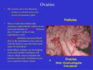
CT basics • Normal Anatomy • Non-neoplasm • Neoplasm Normal Pelvic Anatomy Uterus Ovary
CT basics • Normal Anatomy • Non-neoplasm • Neoplasm Identify structures in next slide:
CT basics • Normal Anatomy • Non-neoplasm • Neoplasm Identify structuresLandmarks: Ovaries usually lateral to uterus and inferior to bifurcation of Iliac vessels C A F E C G D B
CT basics • Normal Anatomy • Non-neoplasm • Neoplasm Identify structures A F E C G D B • Bladder • Piriformis muscle • Right ovary • Rectum • Left ureter • Psoas muscle • Uterine body
CT basics • Normal Anatomy • Non-neoplasm • Neoplasm Another look:
CT basics • Normal Anatomy • Non-neoplasm • Neoplasm Side note: This is a… Hysterosalpingogram Radio-opaque material is injected into the cervical canal. Procedure is used to investigate the shape of uterine cavity and shape and patency of fallopian tubes. Included here to review anatomy
CT basics • Normal Anatomy • Non-neoplasm • Neoplasm Pathology
CT basics • Normal Anatomy • Non-neoplasm • Neoplasm Pathology – Non-neoplasm Describe what you see: • Attenuation (density) • Heterogeneous v. Homogenous • Well-circumscribed v. Indistinct Borders • Enhancing (lights up with contrast) v. non-enhancing • Location
CT basics • Normal Anatomy • Non-neoplasm • Neoplasm Case #1 • 28 year old female presents with fever, lower abdominal pain, new vaginal discharge and complaints of painful intercourse. • Physical exam: Febrile and cervical motion tenderness. • A computed tomography (CT) was done, see next slide.
CT basics • Normal Anatomy • Non-neoplasm • Neoplasm Describe what you see:
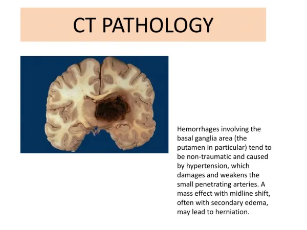
CT basics • Normal Anatomy • Non-neoplasm • Neoplasm Enlarged uterus of soft-tissue attenuation, flanked at the posterior aspects by tortuous, thick-walled oviduct Left greater than Right filled with material of fluid-attenuation. Pelvic Inflammatory Disease
CT basics • Normal Anatomy • Non-neoplasm • Neoplasm Case #2 • 36 year old female presents with sudden onset bilateral pelvic pain, left side worse than right. History of pelvic inflammatory disease a year ago treated with antibiotics. • Physical exam: Tender to palpation in bilateral lower abdomen, L greater than R. Entire pelvis tender to palpation. • A computed tomography (CT) was done, see next slide.
CT basics • Normal Anatomy • Non-neoplasm • Neoplasm Describe what you see:
CT basics • Normal Anatomy • Non-neoplasm • Neoplasm Coronal view - Describe what you see:
CT basics • Normal Anatomy • Non-neoplasm • Neoplasm Left pelvis there is a cystic lesion with heterogeneous enhancement. The lesion appears to be contiguous with the uterus, likely representing… Tubo-ovarian abscess
CT basics • Normal Anatomy • Non-neoplasm • Neoplasm Tubo-ovarian abscess
CT basics • Normal Anatomy • Non-neoplasm • Neoplasm Case #3 • 23 year old female presents with fever, chills, lower abdominal pain, recent history of PID treated with antibiotics. • Physical exam: Febrile and cervical motion tenderness. • A computed tomography (CT) was done, see next slide.
CT basics • Normal Anatomy • Non-neoplasm • Neoplasm Describe what you see:
CT basics • Normal Anatomy • Non-neoplasm • Neoplasm Uterus has large, heterogeneous mass with areas of soft-tissue attenuation and areas of fluid attenuation Tubo-ovarian abscess
CT basics • Normal Anatomy • Non-neoplasm • Neoplasm Case #4 • 23 year old female presents to ED by ambulance due to motor vehicle accident. She is complaining of lower abdominal / pelvic pain. • Physical exam: Pelvis tender to palpation. • A computed tomography (CT) was done, see next slide.
CT basics • Normal Anatomy • Non-neoplasm • Neoplasm Describe what you see:
CT basics • Normal Anatomy • Non-neoplasm • Neoplasm Highly attenuated object in uterus, otherwise normal pelvic CT IUD
CT basics • Normal Anatomy • Non-neoplasm • Neoplasm Case #5 • 21 year old female with 2 days of progressively worsening pelvic pain. She missed last period. She has been feeling nauseated for past 3 weeks. • Physical exam: right pelvic tenderness, breast tenderness. • A computed tomography (CT) was done, see next slide.
CT basics • Normal Anatomy • Non-neoplasm • Neoplasm Describe what you see:
Contrast enhanced axial CT image shows strong enhancing ring-like mass (arrow) that represents gestational sac without hemoperitoneum • CT basics • Normal Anatomy • Non-neoplasm • Neoplasm Ectopic pregnancy
CT basics • Normal Anatomy • Non-neoplasm • Neoplasm Pathology - Neoplasm Describe what you see: • Attenuation (density) • Heterogeneous v. Homogenous • Well-circumscribed v. Indistinct Borders • Enlarged v. atrophied • Enhancing (lights up with contrast) v. non-enhancing • Location
CT basics • Normal Anatomy • Non-neoplasm • Neoplasm Case #6 • 13 year old female presents with abdominal discomfort and feeling bloated. Stomach seems to be growing wider. • Physical exam: Increased abdominal girth • A computed tomography (CT) was done, see next slide.
CT basics • Normal Anatomy • Non-neoplasm • Neoplasm Describe what you see:
CT basics • Normal Anatomy • Non-neoplasm • Neoplasm • Mid pelvis there is large, thin-walled, cystic structure of fluid-attenuation. • At the periphery of this structure are a few distinct regions of heterogeneous tissue. • Within the right lateral aspect lies a foci of bone-density material. • Within the left lateral aspect lies a heterogeneous foci of fat and soft-tissue densities Teratoma (Mature dermatoid cyst)
CT basics • Normal Anatomy • Non-neoplasm • Neoplasm Case #7 • 48 year old female was involved in motor vehicle accident. She is shaken up from accident but otherwise feeling fine. • Physical exam: Pt is in no acute distress. No signs or symptoms of pain. Patient insisted having a CT to rule out bleeds. • A computed tomography (CT) was done, see next slide.
CT basics • Normal Anatomy • Non-neoplasm • Neoplasm Describe what you see: Clue: this arises from the ovary
CT basics • Normal Anatomy • Non-neoplasm • Neoplasm Posterior aspect of the pelvis lies a well-circumscribed, thin-walled, non-septated cystic structure containing fluid-density material Clue: this arises from the ovary Serous Cystadenoma (benign) Incidental finding on CT
CT basics • Normal Anatomy • Non-neoplasm • Neoplasm Case #8 • 58 year old female presents to clinic with bloating, back pain, urinary urgency, constipation, and tiredness for 6 months. Recently she developed pelvic pain, vaginal bleeding, and unintentional weight loss. • Physical exam: Abdomen tender to palpation throughout. Pelvic tenderness. • A computed tomography (CT) was done, see next slide.
CT basics • Normal Anatomy • Non-neoplasm • Neoplasm Describe what you see: Clue: This arises from the ovary
CT basics • Normal Anatomy • Non-neoplasm • Neoplasm The pelvic cavity is grossly distended by multiple well-circumscribed, thin-walled, septated, lobular structures of fluid-density. These structures are compressing but don’t seem to invade surrounding pelvic tissues Cystadenocarcinoma (Malignant) Note: Septations and lobulatedsurface
CT basics • Normal Anatomy • Non-neoplasm • Neoplasm Sources Siddall KA. Multidetector CT of the female pelvis. RadiolClin North Am. 01-NOV-2005; 43(6): 1097-118 Casillas J, Joseph RC, Guerra JJ Jr. CT appearance of uterine leiomyomas. Radiographics. 1990 Nov;10(6):999-1007. Foshager MC, Walsh JW. CT anatomy of the female pelvis: a second look. Radiographics. 1994 Jan;14(1):51-64; Outwater EK, Siegelman ES, Hunt JL. Ovarian teratomas: tumor types and imaging characteristics. Radiographics. 2001 Mar-Apr;21(2):475-90. Pannu, HK, et al. MD CT Evaluation of Cervical Cancer: Spectrum of Disease. Radiographics 2001; 21:1155–1168 Rha SE, et al. CT and MR imaging features of adnexal torsion. Radiographics. 2002 Mar-Apr;22(2):283-94. Roberts JL, Dalen K, Bosanko CM, Jafir SZ. CT in abdominal and pelvic trauma. Radiographics. 1993 Jul;13(4):735-52. Roobolamini, SA. Imaging of Pregnancy-related Complications. Radiographics 1993; 13:753-770. Saksouk FA, Johnson SC. Recognition of the ovaries and ovarian origin of pelvic masses with CT. Radiographics. 2004 Oct;24 Suppl 1:S133-46. Sam JW, Jacobs JE, Birnbaum BA. Spectrum of CT findings in acute pyogenic pelvic inflammatory disease. Radiographics. 2002 Nov-Dec;22(6):1327-34. Yang DM. Retroperitoneal cystic masses: CT, clinical, and pathologic findings and literature review. Radiographics. 2004 Sep-Oct;24(5):1353-65. Buy, J-N, et al. Cystic Teratoma of the Ovary: CT Detection. Radiology 1989; 171:697-701 As well as IMPAX, EPIC, and WIKIPEDIA