Download
1 / 70
830 likes | 1.77k Views
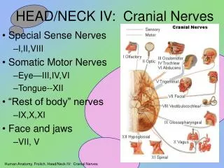
Anatomy for PET/CT Head and Neck . Dr Jagrit Shah Consultant Neuroradiologist & Head and Neck Radiologist Nottingham University Hospitals NHS Trust. Contents. Neck Nodes Neck spaces Mucosal lesion location Some examples. Objectives. By the end of this talk you should be able to

E N D
Anatomy for PET/CTHead and Neck Dr Jagrit Shah Consultant Neuroradiologist & Head and Neck Radiologist Nottingham University Hospitals NHS Trust
Contents • Neck Nodes • Neck spaces • Mucosal lesion location • Some examples
Objectives • By the end of this talk you should be able to • Confidently localise a PET/SPECT positive lesion when reporting lesion in the Head and Neck • Teach others basic Head and Neck anatomy
Question • What is the best way of determining the mucosal extent of squamous cell carcinoma within the Head and Neck? • MRI scan • CT scan • PET/CT • Get on the phone