Download
1 / 16
160 likes | 275 Views
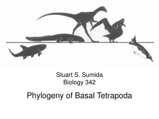
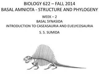
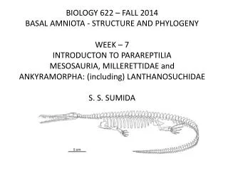
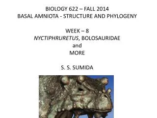
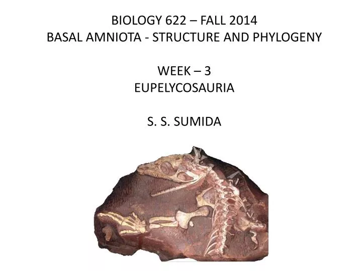
BIOLOGY 622 – FALL 2014 BASAL AMNIOTA - STRUCTURE AND PHYLOGENY WEEK – 3 EUPELYCOSAURIA S. S. SUMIDA.

E N D
BIOLOGY 622 – FALL 2014 BASAL AMNIOTA - STRUCTURE AND PHYLOGENY WEEK – 3 EUPELYCOSAURIA S. S. SUMIDA
Recall the Synapsida is one of the two great groups of the Amniotasensustricto (the other being Reptilia). Within Synapsida, two subgroups are known, the Caseasauria and the Eupelycosauria. The term “Eupelycosauria” has been adopted in part as a reaction to the realization that “Pelycosauria” is technically a polyphyletic group, as it does not include all members of the group derived from it. So, notably, Eupelycosauria technically includes all therapsids and mammals as well. We are focusing only the basal-most members. As stated earlier, the familial designations of Romer and Price (1940) have in many cases held up, however, the interrelationships of those families have been subject to some change.
MODERN INTERPRETATIONS OF PHYLOGENETIC RELATIONSHIPS OF BASAL SYNAPSIDS (I.E “PELYCOSAURS”) The work of Reisz (1980, 1986), Reisz et al (1992), and Berman et al (1995) have confirmed the validity pelycosaurian families, and placed them in two distinct groups: SYNAPSIDA Caseasauria Family Eothyrididae Family Caseidae Eupelycosauria Family Varanopseidae Family Ophiacodontidae Family Edaphosauridae Sphenacodontia Haptodus Palaeohatteria Pantelosaurus Cutleria Sphenacodontoidea Family Sphenacodontidae Therpasida(leading to mammals)
EUPLEYCOSAURIA: DEFINITION Eupelycosauria is defined as those taxa bearing all basal synapsidsynapomorphies with the following unique features: (1) a long, narrow supratemporal. (2) The frontal contributes to at least one third of the dorsal orbital margin. Haptodus, Palaeohatteria, Pantelosaurus, and Cutleria are all genera that used to be placed in a distinct group known as the “Haptodontidae” or Haptodontinae”; but that has been shown to be a structural grade as opposed to a monophyletic family or subfamily.
THE MAJOR GROUPS OF EUPELYCOSAURIA OVERVIEW: What seems clear is that a few features are common to all eupelycosaurs, but as Romer determined as far back as 1940, there are certain families that have been robust in terms of their stability over many years.
Defining Features of EUPELYCOSAURIA • A long, narrow supratemporal. The supratemporal of caseasaurs is almost as wide as long (length to width ratio less than two). • 2) The frontal contributes to at least one third of the dorsal orbital margin (the orbital contribution of the frontal is narrower in caseasaurs). • In this case a varanopid skull provides a basal example of a eupleycosaur to show these features.
Defining Features of VARANOPSEIDAE Following is one of the most famous specimens of a varanopid, Aerosaurus from the Early Permian of northern New Mexico.
Defining Features of VARANOPSEIDAE • Marginal dentition composed of strongly curved, mediolaterally flattened teeth. • Occipital flange of squamosal reduced. • Narrow zygomatic arch.
Defining Features of SPHENACODONTIA • Maxillary supracanine buttress present (this structure is a thickening of the maxilla visible on its internal surface, dorsal to the caniniform teeth). • Premaxillary teeth in deep sockets. The teeth of other synapsids are implanted in shallow sockets. • Example is from Dimetrodon.
The “HaptodontineGrade” of Organization Haptodus and the genera usually considered to be closely related to it have traditionally been lumped together in the family Haptodontidae or subfamily Haptodontinae. However, recent work has suggested they instead represent a series of small transitional genera between Sphenacodontines and more basal eupelycosaurs.
Defining Features of SPHENACODONTOIDEA • Frontal orbital process extends far laterally (it is poorly developed, if present, in other synapsids). • Deep prefrontal pocket (a concavity near the anterodorsal edge of the orbit). • Vomerine teeth absent (the vomer bears a shagreen of denticles in other synapsids).
Defining Features of SPHENACODONTIDAE • Presence of a ventral narial process of the nasal (just posterior to the external naris) • Frontal anterior process narrower than its posterior process (these two processes are equally wide in other synapsids). • Postorbital-squamosal contact extensive (this contact is narrow in other synapsids, when present) . • Supratemporal contacting postorbital (this contact is absent in most other eupelycosaurs). • Paroccipital process extends ventrolaterally and posteriorly (it extends laterally in most other synapsids). • Caniniform root bulges into choana (in other synapsids, the caniniform tooth does not constrict the choana).
Defining Features of SPHENACODONTIDAE • Presence of a ventral narial process of the nasal (just posterior to the external naris) • Frontal anterior process narrower than its posterior process (these two processes are equally wide in other synapsids). • Postorbital-squamosal contact extensive (this contact is narrow in other synapsids, when present) . • Supratemporal contacting postorbital (this contact is absent in most other eupelycosaurs). • Paroccipital process extends ventrolaterally and posteriorly (it extends laterally in most other synapsids). • Caniniform root bulges into choana (in other synapsids, the caniniform tooth does not constrict the choana).