Download
1 / 8
80 likes | 308 Views
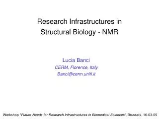
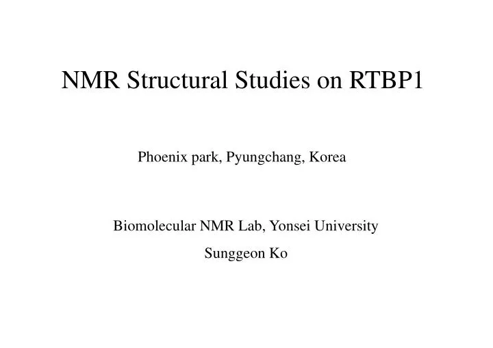
NMR Structural Studies on RTBP1. Phoenix park, Pyungchang, Korea. Biomolecular NMR Lab, Yonsei University Sunggeon Ko. 500. 600. 633. 400. 300. 100. 1. 200. N-. -C. 506. 615. Myb-like domain. GST-.

E N D
NMR Structural Studies on RTBP1 Phoenix park, Pyungchang, Korea Biomolecular NMR Lab, Yonsei University Sunggeon Ko
500 600 633 400 300 100 1 200 N- -C 506 615 Myb-like domain GST- 506PFADP511NSLAL 516ANVPL 521SRSKR 526PDFGQ 531RRIRR 536PFTVA 541EVELL 546VEAVE 551HLGTG 556RWRDV 561KFRAF 566ENVHH 571RTYVD 576LKDKW 581KTLVH 586TASIA591PQQRR 596GAPVP 601QELLD 606RVLAA 611QAYWS < 110 amino acid sequence (11.5kD) of the DNA binding domain of RTBP1 > The DNA binding domain of RTBP1(Rice Telomere Binding Protein) • Protection of the end of chromosome from recombination, fusion and recognition as damaged DNA • - Characteristics of specific binding to tandem repeats of rice telomere (GGGTTT) • Duplex DNA binding protein • The Myb-like domain acts as a homodimmer whereas the Myb-like domain of TRF1 does as a monomer A B Yonsei Biomolecular NMR Lab
Sequential alignment of the DNA binding domains A) B) hTRF1 : Human telomere binding protein (Homo sapiens) RTBP1 : Rice telomere binding protein 1(Oryza sativa) ngTRF1 : Telomere repeat binding factor (Nicotiana glutinosa) atTRF1 : Telomere repeat binding factor (Arabidopsis thaliana) Yonsei Biomolecular NMR Lab
Bindign activity to telomere repeat (TTTAGGGTTTAGGG) Myb 1 586 633 532 RTBP1 + 506 596 532 615 RTBP1506-615 - 506 532 594 RTBP1506-594 - 528 615 596 RTBP1528-615 - 528 594 Sequence RTBP1528-594 Telomere two repeats RTBP fragment 1 506-615 RTBP1528-594 RTBP1528-615 RTBP1506-615 RTBP1506-594 Free probe RTBP fragment 2 506-594 200ug 200ug 400ug 200ug 200ug 200ug 200ug 400ug 400ug 400ug 800ug 800ug 800ug 800ug 528-615 RTBP fragment 3 528-594 RTBP fragment 4 5’ TTTAGGGTTTAGGG 3’ AAATCCCAAATCCC Characterization of the DNA binding domain by EMSA B ) A) C ) Characterization of the DNA binding domain of RTBP1 -Telomere DNA repeats in plants(A) and recombinant fragments of the DNA binding domain of RTBP1(B) and EMSA assay of each fragments (C) Yonsei Biomolecular NMR Lab
NMR experiments and structure calculation 1. Sample preparation and NMR experiment - All NMR samples contained 100mM NaCl and 1mM NaN3 in 50mM potassium phosphate at pH 7.0. Measurements were done in mixture of 90% 1H2O and 10% 2H2O and 99% 2H2O as solvent by Bruker DRX-500MHz, a Varian Unity INOVA 500MHz spectrometer or a Bruker DRX-800MHz equipped with a triple resonance probe with x, y, z-gradients in 303K 2. NMR spectra and constraints of structure calculation - Sequential assignment : 15N-edited TOCSY, 15N-edited NOESY, 2D-HSQC, HNCO, HNCA, HNCACB and CBCA(CO)NH - Side chain assignment : 15N-edited TOCSY, 15N-edited NOESY, HCCH-TOCSY and 13C-edited NOESY - NOE assignment: 15N-edited NOESY and 13C-edited NOESY - Angle constraint : HNHA and Talos - Hydrogen bond : Slowly exchanging amide protons were identified by lyophilization of a fully protonated sample in H2O solution to dryness, re-dissolution in a 99.99% 2H2O solution, and immediate acquisition of a two-dimensional 1H-15N HSQC spectra or acquisition of it in a day - Spin relaxation measurements : Heteronuclear cross-relaxation rates (NOEs) was acquired by interleaving pulse sequences with and without proton saturation. 3. Structure calculation - Calculation : CNS 2.0 on SGI Indigo2 workstation - Display and analysis : MOLMOL and Insight II Yonsei Biomolecular NMR Lab
T554 F507 H585 Q530 D527 E547 V584 E543 L515 V113 A587 (N567) Y613 A548 L545 V549 K561 (Q601) R556 (Q593) F528 V542 G555 K579 D509 W580 G596 H552 A540 V518 (N511) G528 A609 R532 (Q611) G117 R595 (Q530) S588 L520 G553 I533 R532 V599 (N567) A610 A516 (N517) D114 (Q582) L513 H552 V607 R118 (N511) T582 N567 Q586 A514 (Q530) I119 V560 W614 T572 S615 S512 F565 T538 R535 K579 T586 E602 R563 S615 A564 L552 E542 Q593 N511 L608 N517 S521 Y573 L603 L544 L576 Q612 F562 T121 V546 S116 I589 H585 R571 D578 E547 D605 L101 F507 D559 V568 E543 V574 R534 E550 V113 D575 K577 R606 V539 R558 V518 F537 R525 A597 W557 A508 V120 A540 Q601 R534 E566 L593 R522 W614 W557 W580 1H-15N HSQC Spectra of the DNA binding domain Ko, S.G.,et al,.(2003) J.Biomol NMR, Sep, 27(1):89-90 The 2D 1H-15N HSQC spectrum for the DNA binding domain in RTBP1 labeled with assignments. -Unlabeled peaks do not indicate the residues of the DNA binding domain in RTBP1 that has only 109 amino acids, but the residues of the vector, p-GEX 4T-1, that adds 10 amino acids to C-terminal of the DNA binding domain of RTBP1. Yonsei Biomolecular NMR Lab
540 C 40 – 45 - 50 - 55 - 60 - 65 - 548 C • 20 • 30 • 40 • 45 • 50 • 55 • 60 • 40 • 45 • 50 • 55 • 60 • - 65 543 C 547 C 542 C 539 C 546 C 541 C 544 C 545 C 548 C 540 C 544 C 545 C 547 C 538 C 543 C 541 C 539 C 538 C 546 C 542 C 538T 539V 540A 541E 542V 543E 544L 545L 546V 547E 548A 542V 543E 544L 545L 546V 547E 548A 549V 550E 551H 552L 553G 554T 555G 556R 557W Sequential assignment of RTBP1 A) B) Sequential assignment using HNCA(A) and HNCACB(B) contained CBCA(CO)NH, HNCO Yonsei Biomolecular NMR Lab
Conclusion 1. The DNA binding domain of hTRF1 binds as a monomer and the Myb-like domain , only three helixes, of protein is enough to make DNA-protein complex. (Nishikawa et al., (2001) Structure, 9, 1237-1251) 2. The DNA binding domain of RTBP1 binds as dimmer and needs extra secondary structures, helix I and helix V, confirmed by NMR 1D-titration and EMSA assay. 3. In the result of the sequential alignment of the DNA binding domain in hTRF1 and RTBP1, they have a high sequence homology(31%) but the solution structure between them is very different which explains why each proteins have a different DNA binding mode. 4. Most plants have a same telomere repeats (TTTAGGG) and very similar amino acids in telomere binding factors, especially in C-terminal. And most of plant telomere proteins, RTBP1, ngTRF1 and AtTBP1, needs a Myb-like domain and extra amino acid sequences especially positive charged Amino acids in pH7, in N-and C-terminal. Dynamics study, although preliminary data, and the distribution of positive charged amino acids on surface of RTBP1 shows that RTBP1 needs additional N-, C-terminal, helix I, helix IV-V ioop and helix V, to make a plant telomere. 5. Although plant and human have a similar DNA binding motif, a Myb-like domain, but they use a different mechanism to make a telomere which means they have a same origin but are evolved by special environments and each purposes that are remained us to identify. Yonsei Biomolecular NMR Lab