Download
1 / 20
200 likes | 204 Views
Learn how tandem MS analysis overcomes the limitations of peptide mass identification and allows for accurate protein identification based on peptide sequencing. Explore the principle, fragmentation process, and different types of peptide fragments in MS/MS analysis.

E N D
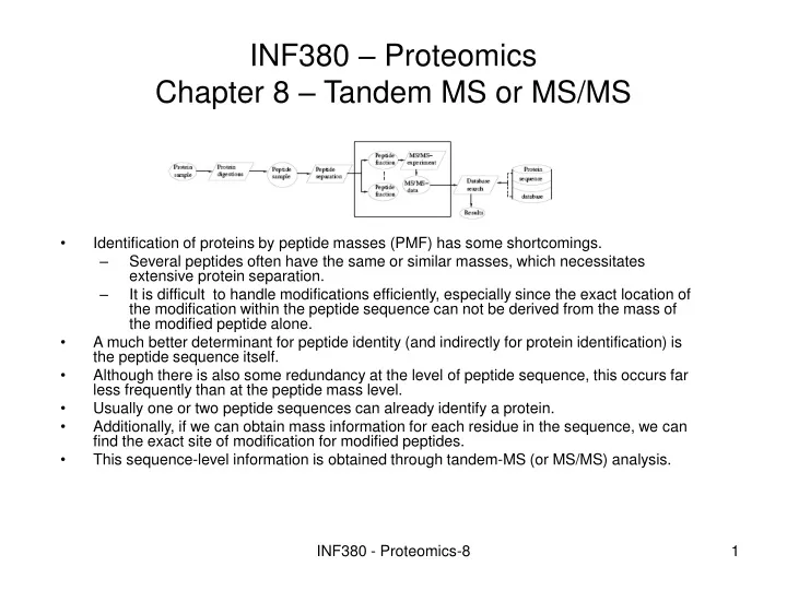
INF380 – Proteomics Chapter 8 – Tandem MS or MS/MS • Identification of proteins by peptide masses (PMF) has some shortcomings. • Several peptides often have the same or similar masses, which necessitates extensive protein separation. • It is difficult to handle modifications efficiently, especially since the exact location of the modification within the peptide sequence can not be derived from the mass of the modified peptide alone. • A much better determinant for peptide identity (and indirectly for protein identification) is the peptide sequence itself. • Although there is also some redundancy at the level of peptide sequence, this occurs far less frequently than at the peptide mass level. • Usually one or two peptide sequences can already identify a protein. • Additionally, if we can obtain mass information for each residue in the sequence, we can find the exact site of modification for modified peptides. • This sequence-level information is obtained through tandem-MS (or MS/MS) analysis. INF380 - Proteomics-8
Principle for MS/MS INF380 - Proteomics-8
Principle for MS/MS • The principle for MS/MS analysis relies on two mass analyzers in tandem (hence the name). • The first mass analyzers allows the selection of a particular m/z range, usually centered on the m/z of a peptide of interest. • The selected precursor subsequently undergoes a fragmentation step with the second mass analyzer finally measuring the m/z of the resulting fragment ions. • The resulting spectrum, which contains the m/z and ion intensity for each of the fragment ions is called an MS/MS spectrum • A more standardized nomenclature refers to a fragmentation spectrum as a product ion spectrum. INF380 - Proteomics-8
Principle for MS/MS • For identification and characterization the MS/MS-spectrum is compared to subsequences in a database, by using one of the approaches described earlier • The peptide ion chosen for fragmentation is called precursor ion or parent ion • The resulting fragment ions are likewise called product ions or daughter ions. • Fragments (or fragment parts) that are neutral are called neutral losses. • Note that the presence of these neutral fragments can only be inferred by observing both the original fragment with and without the loss. They can not be seen directly due to the lack of charge. INF380 - Proteomics-8
Peptide fragments • The peptides are mainly fragmented along the peptide backbone. • Further fragmentation may occur, changing the produced fragments into other fragment types. • Since the first mass analyzer selects ions that fall within a certain m/z value rather than a single m/z value, multiple ions can be selected simultaneously. • Each of these selected precursors will yield fragments, of which those with sufficiently high abundances are shown as peaks in the MS/MS-spectrum. • The precursor can carry one or more charges, depending on the ionization source and the peptide properties. INF380 - Proteomics-8
Peptide fragments • When a singly charged precursor ion fragments, the single charge will necessarily be located on only a single fragment ion, effectively hiding the neutral sister fragment from the mass spectrometer. • With multiply charged precursors, the different charges are usually distributed across the product ions. • One advantage with multiply charged precursor is that instruments with lower maximum precursor m/z values can be used. • Multiply charged ions also tend to fragment easier, and as explained above, yield more fragment ions per fragmentation event. • The accepted nomenclature for the fragment ions is to denote each type with a letter. INF380 - Proteomics-8
Peptide fragments • Backbone fragments result from fragmentation along the peptide backbone. • If some charge is retained on the N terminal fragment, the fragment ion is classified as either a, b or c, depending on which bond is broken. • If some charge is retained on the C terminal fragment, the fragment type is either x, y or z. • The fragment ions are annotated by subscripts, indicating the number of residues in them • Internal fragments result from double backbone fragmentation. Usually, these are formed by a combination of b type and y type fragments, and consist of five residues or less. INF380 - Proteomics-8
Peptide fragments • Immonium fragments are internal fragments composed of a single side chain formed by a combination of a a type and y type fragmentation. • This means that both a C and O atom (-28 Da) are lost compared to the residue, see Figure • However, it abstracts an extra proton, hence an immonium ion for an amino acid is observed at a m/z 27 Da less than the residue mass of the amino acid (assuming single charge). • A peak (with sufficient abundance) at the mass of an immonium ion (strongly) indicates the presence of the corresponding amino acid. • Water loss occurs when backbone fragments lose a water molecule (H2O, -18.011 Da). • The resulting fragments are denoted ao, bo etc. • Ammonia loss occurs when fragments lose an ammonia molecule (NH3, - 17.027 Da). • The resulting fragments are denoted a* etc. • Side chain fragmentation occurs when additional fragmentation of the side chain of a backbone fragment takes place. The most common are • d-ions from partial side chain fragmentation of a-ions, • v-ions from complete side chain fragmentation of y-ions • w-ions from partial side chain fragmentation of z-ions. • Side chain fragmentation can be used to differentiate between leucine and isoleucine, since the resulting fragments will have different masses. INF380 - Proteomics-8
Peptide fragments INF380 - Proteomics-8
Fragmentation techniques • CID – Collision-Induced Dissociation is the most common technique for fragmentation INF380 - Proteomics-8
MS/MS spectrometers and analyzers • There exist a lot of different analyzers for MS/MS • One of them is TOF/TOF INF380 - Proteomics-8
Overview of the process for MS/MS-analysis • An MS-spectrum is first (conceptually) constructed for every spot location (MALDI) or at given time points for an ESI instrument • From this measurement, the peaks for subsequent MS/MS-processing are automatically selected.The criteria for this selection can usually be user-specified: • the n most intense peaks, n normally between three and eight • charge state • An inclusion list with the m/z values to analyze can sometimes also be specified. INF380 - Proteomics-8
Fragment ion masses and residue masses • Interpretation of the spectra relies on knowing the relation between the mass of a fragment type, and the sum of the residue masses of the amino acids in the fragment ion. INF380 - Proteomics-8
Fragment ion masses and residue masses • <N> the (nominal) mass of the N-terminal group, = 1 (H). • <C> the (nominal) mass of the C-terminal group, = 17 (OH). • RP the sum of the residue masses of a peptide under consideration. • MP the neutral mass of a peptide under consideration (=RP+<N>+<C>). • RF the sum of the residue masses of a fragment ion under consideration. • Mk the mass of a fragment ion of type k under consideration (charge one). • The masses for the singly charged fragment ion types can now be written as: • Mb=RF+<N> My=RF+<C>+2H • Ma=RF+<N>-CO Mx=RF+<C>+CO • Mc=RF+<N>+N+3H Mz=RF+<C>-NH INF380 - Proteomics-8
Complementary masses • Adding complementary masses results in(note that RP is the sum of RF for the two complementary fragment types) • Mb+My=RP+<N>+<C>+2H (=MP+2H) • Ma+Mx=RP+<N>-CO+<C>+CO (=MP) • Mc+Mz=RP+<N>+N+3H+<C>-N-2H (=MP+2H) • The most frequently occurring fragment types by far for CID are the b- and y-ions and partly a-ions, and many programs consider only these. We will also mainly consider these here for illustration purposes. INF380 - Proteomics-8
Deisotoping and charge state deconvolution • One goal of preprocessing is to obtain a spectrum where each fragment is represented by only one datum, which means that • an isotope envelope is reduced to one peak (deisotoping), • a fragment that occurs in different charge-states is also collated into one state (commonly to the singly charged ion, which is called charge state deconvolution). • An m/z for z>1 is converted to [((m/z)-1)z+1], and the intensities are summed for ions occurring with several charges • These two tasks both rely on the m/z differences between the peaks, and can therefore be done simultaneously. • A serious problem is that isotopic envelopes can overlap, causing the intensity of a single, compound peak to be split over two peaks in the deisotoped and deconvoluted spectrum. INF380 - Proteomics-8
Deisotoping and charge state deconvolution • Programs for charge state determination typically define a maximum limit for the charge (three or four for fragment ions). • Furthermore, the charge can not be higher than the charge of the precursor, and for each fragment it can not be so large that the neutral mass of the fragment becomes larger than the mass of theprecursor. • For each peak, the occurrence of isotopes is checked against the predicted masses of the suggested charge. • One can also take the expected number of isotopes for themass of the peak into account, to see if these exist. • When it is found that a peak may belong to more than one isotopic envelope, the problem of distributing the intensity of that peak occurs. • A reasonable way of solving this is to use modelsfor the expected intensity distribution of the isotope envelopes included. INF380 - Proteomics-8
Precursor mass correction • Precursor mass errors can occur when the wrong isotope has been selected as the monoisotopic peak, or if the charge is either not determinable, or incorrectly determined. • Precursor mass errors can be controlled for or estimated from their MS/MS spectrum by using complementary fragment ions • We show the principle assuming that the precursor charge is two. • For complementary ions (b,y) and (c,z) we have the equation Mb+My=MP+2H. • Let R=r1,...,rm be a spectrum. • A reversed spectrum is defined as R*=r*1,...,r*m, where r*i=MP+2H-ri, • Then b(c)-ions in R should be translated to corresponding y(z)-ions in R*, and y(z)-ions to b(c)-ions. • There should therefore be many common peaks in R and R*. • If MP is not correct however, this correspondence will not occur. • MP can therefore be considered an unknown x, which should be determined. • Then define R*(x) with r*i=x+2H-ri. • Let then C(x)=C[R,R*(x)] be the number of common peaks (to a given accuracy) in R and R*(x). • The value of x that maximizes C(x) should be chosen as the corrected precursor mass. • This problem can be solved as a mixed integer programming problem, but also in polynomial time (O(m3)) by a straightforward algorithm INF380 - Proteomics-8
Estimating the charge state of the precursor • While MALDI rarely produce ions of charge state higher than one, multiple charge ions are commonly produced by ESI. • However, fragmentation spectra from precursors with a charge higher than three are seldom of good enough quality for subsequent identification. • The problem is therefore reduced to determining the charge as being either one, two, three or ``larger''. The charge state is commonly found by using the isotopic differences of the associated MS-spectrum. • However, especially for low resolution instruments, the charge state assignment may be wrong or simply unknown. • Knowing the actual charge state is important for performing a reasonable database search, all the more so since erroneous charge state assignments often lead to faulty results. • An alternative to determining the correct charge state of the precursor is to perform the database for several states, but this is time consuming, and it is not always possible to select the correct search results afterwards. • Several methods have therefore been developed for estimating the precursor charge state from MS/MS-spectra. • The easiest approach is to differentiate between charge one and greater than one. • If the charge is one, the (m/z) of all the product ions are less than (m/z) of the precursor. For example let T+ be the total ion current for ions with (m/z) greater than that of the precursor, and T- the rest total ion current. If the ratio (T+/T-) is small (for example less than 0.1), the charge state can be estimated to one. • 2to3 is a program that estimates the charge (2 or 3) of multiple charged precursors. INF380 - Proteomics-8
MS3 spectra • Since the analysis in in-time analyzers uses the same analyzers for MS and MS/MS, one can actually repeat the process. • Product ions from MS/MS can be selected for another fragmentation step and an MS3 spectrum is thus produced. • The process can be repeated even more, producing MSn spectra, for n-1 fragmentation steps. • The number of ions available for fragmentation will decrease, such that a limit for producing reliable spectra from today's instruments is reached for an n of about ten. • MS3 spectra can be used to resolve ambiguities in an identification. • For identification MS/MS spectra are compared to segments in a protein sequence database. • A segment is a subsequence, in our context often a theoretical peptide. • When more than two segments can fit in an MS/MS spectrum, the most intense peak in the spectrum can be selected for a second fragmentation step and the resulting MS3 spectrum can then be compared to the two candidate segments. • It can also be useful when no matching segment is found for the MS/MS spectrum, since the MS3 spectrum is simpler and may be easier to reveal helpful information. INF380 - Proteomics-8