Download
1 / 28
280 likes | 420 Views
T Cell Activation/Function Ned Braunstein, MD ned_braunstein@merck.com nsb2@columbia.edu. The Major T Cell Subsets. CD4+ T cells. CD8+ T cells. lck. p56. lck. p56. z. z. z. z. g. g. d. d. e. e. CD3. CD3. C. C. a. b. C. C. a. b. CD8. TCR. TCR. V. CD4. b. V. b.

E N D
T Cell Activation/Function Ned Braunstein, MD ned_braunstein@merck.com nsb2@columbia.edu
The Major T Cell Subsets CD4+ T cells CD8+ T cells lck p56 lck p56 z z z z g g d d e e CD3 CD3 C C a b C C a b CD8 TCR TCR V CD4 b V b V a V a peptide peptide MHC I MHC II (1) Interacts with MHC class II expressing cells (B cells, macrophages) (2) Induce(help) B cells to synthesize antibody (3) Induce and activate macrophages (4) Secretes lymphokines (1) Interacts with MHC class I expressing cells (all nucleated cells) (2) Kill MHC class I expressing target cells (3) Suppress immune responses (4) Secretes lymphokines
Naïve T cells are activated by antigen in LNs where they mature into effector cells (1) Naive T cells home continuously from the blood to lymph nodes and other secondary lymphoid tissues. Homing to lymph nodes occurs in high endothelial venules (HEV), which express molecules for the constitutive recruitment of lymphocytes. (2) Lymph fluid percolates through the lymph nodes; the fluid is channeled to them from peripheral tissues, where dendritic cells collect antigenic material. In inflamed tissues, dendritic cells are mobilized to carry antigen to lymph nodes, where they stimulate antigen-specific T cells. On stimulation, T cells proliferate by clonal expansion and differentiate into effector cells, which express receptors that enable them to migrate to sites of inflammation. (3) Although most effector cells are short-lived, a few antigen-experienced cells survive for a long time. These memory cells are subdivided into two populations on the basis of their migratory ability: the effector memory cells migrate to peripheral tissues, whereas central memory cells express a repertoire of homing molecules similar to that of naive T cells and migrate preferentially to lymphoid organs.
The Biology of Dendritic Cells: Antigen capture and presentation to T cells Antigens are captured by DCs in peripheral tissues and processed to form MHC-peptide complexes. As a consequence of antigen deposition and inflammation, DCs begin to mature, expressing molecules that will lead to binding and stimulation of T cells in the T-cell areas of lymphoid tissues. If the antigen has also been bound by B cells, then both B and T cells can cluster with DCs. After activation, B blasts move to the lining of the intestine, the bone marrow, and other parts of the lymphoid tissue with some becoming antibody-secreting plasma cells. T blasts leave the blood at the original site of antigen deposition, recognizing changes in the inflamed blood vessels and responding vigorously to cells that are presenting antigen. This limits the T-cell response to the site of microbial infection. Banchereau, J. and Steinman, R. Nature 392, 245 - 252 (1998)
The T cell activation cycle Minutes Hours Days IL-2R P P Ca++ IP3 DAG c- fos IL-2 etc. c- jun Antigen recognition c- myc NF-KB Cytokine production and autocrine stimulation NF-AT Immediate events Effector functions: Help DTH Killing (CTL) regulation Proliferation
PKC MHC + Ag ICAM-1 (CD54) CD80 CD4 LFA-1 (CD11a/CD18) CD3 CD28 IL2R lck fyn CD45 ZAP-70 PTK RAS Cytoplasm PLC PIP2 DAG IP3 MAP-kinase Ca Nucleus IL-2 NF-AT NF-KB OTF1
PKC MHC + Ag CD80 ICAM-1(CD54) CD4 LFA-1(CD11a/CD18) CD3 CD28 IL2R lck fyn CD45 ZAP-70 PTK RAS Cytoplasm PLC PIP2 DAG IP3 MAP kinase Ca2+ I-kB P NF-kB Nucleus IL-2 NF-AT NF-kB OTF1
CNB CNA Cal- PKC modulin MHC + Ag CD80 ICAM-1 (CD54) CD4 LFA-1(CD11a/CD18) CD3 CD28 IL2R lck fyn CD45 ZAP-70 PTK Cytoplasm PLC PIP2 DAG IP3 Ca2+ P NF-AT Nucleus Fos Jun P IL-2 NF-AT NF-kB OTF1
The T cell activation cycle Minutes Hours Days IL-2R P P Ca2+ IP3 DAG c- fos IL-2 etc. c- jun Antigen recognition c- myc NF-kB Cytokine production and autocrine stimulation NF-AT Immediate events Effector functions: Help DTH Killing (CTL) regulation Proliferation
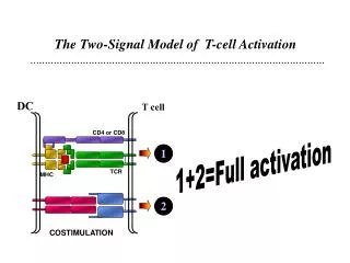
Key molecular interactions between T cells and APCs MHC class II/ autopeptide CD3 TCR CD40 CD80 Activated T cell CD40 CD40L CD40 CD28 CD80 MHC class II (1) induction of cytokines/chemokines (IL-8, IL-12, TNF-a, MIP-1a) (2) stimulation of CD80 and CD86 expression and co-stimulatory function with activation of T cell growth (3) augmentation of antigen-presenting function
Naïve CD4+ T cells differentiate into Th1 and Th2 subsets Th1 Cells IL-2IFN-g TNF IL-2 IFN-g, IL-12 Antigen + APC IFN-g (–) IL-4 IL-10 (–) Resting CD4+ cell “pTh” Activated CD4+ cell IL-4 IL-5 IL-6 IL-10 IL-4, IL-13 Th2 Cells
Functions of Th subsets Functions of Th1 subsets Th1 Cells • Activate macrophages/dendritic cells augment antigen presentation • induce delayed type hypersensitivity (DTH) responses important in eradicating intracellular pathogens (TB, leprosy, listeria • mediate Th1 diseases (ie; rheumatoid arthritis, multiple sclerosis and type I diabetes IL-2IFN-g TNF IL-4 IL-10 (–) IFN-g (–) Functions of Th2 subsets IL-4 IL-5 IL-6 IL-10 • Help B cells and induce humoral immunity • mediate allergic and immediate hypersensitivity responses • involved in antibody mediated immune diseases like SLE and ITP Th2 Cells
Major Functions of T Lymphocytes (1) Induction and Activation of B cells (Help)-required for most antibody responses (2) Delayed Type Hypersensitivity (DTH) - important in elimination of intracellular pathogens (virus, fungi and mycobacteria) (3) Cell mediated Cytotoxicity (Killer function)-important in the immune response to virus infected cells and cancer cells (4) Suppressor Cell Function- regulates the cell mediated and antibody responses
The Induction and Activation of B cells (Helper Function) Antigen binds specifically to SmIg, is internalized into vesicles and cleaved into peptides which displace CLIP and bind to MHC class II molecules in the endocytic compartment. The peptide/MHC complex is then transported to the surface membrane. The CD4+ T cell a,b TCR/CD4 complex binds to the MHC class II/peptide complex on the surface of B cells. The CD4+ Th2 cells are triggered to secrete IL-2, IL-4, IL-5, IL-6 and IL-10 and begin to express CD40L. These lymphokine and contact dependent signals (CD40L) induce B cells to proliferate, class switch and differentiate into antibody (IgM, IgG, IgA and IgE) secreting B cells and plasma cells.
Antigen Processing and Presentation by B cells ANTIGEN MHC Class ll BCR (SmIg) B cell Peptide Internalization of antigen/Ig Antigenic peptides Bind to MHC class II molecules Antigen binds specifically to BCR (surface membrane Ig), is internalized into vesicles and cleaved into peptides which displace and bind to MHC class II molecules. The peptide/MHC complex is then transported to the surface membrane.
Antigen Presentation by B cells BCR (SmIg) hapten Antigen MHC class II carrier protein MHC class II/ carrier peptide complex B cell carrier peptides Antigen binds specifically to SmIg, is internalized into vesicles and cleaved into peptides which displace and bind to MHC class II molecules. The peptide/MHC complex is then transported to the surface membrane.
Expression of Membrane Proteins Following Antigen Specific Activation of T and B Cells BCR (SmIg) TCR Resting EffectorT cell Resting B cell MHC class II CD4 BCR (SmIg) CD23 IL-2R TCR Activated Effector T cell CD40 CD40L CD4 MHC class II CD80 CD86 Activated B cell MHC class II
CONSEQUENCES OF CD40L/CD40 INTERACTIONS DURING T-B CELL INTERACTIONS CD23 CD40L TCR Sm Ig CD4 CD40 Activated Effector T cell ActivatedB Cell • Triggering of B cell proliferation • Rescue from apoptosis • Induction of Ig isotype class switching • Up-regulation of CD80 and CD86 • Germinal center formation • Up-regulation of CD23 • Downregulation of CD40L expression
CD23 SmIg CD40 Activated B cell IgG IgA IgE Plasma Cell Final Phases of B cell Differentiation are Mediated by Contact T cell signals (CD40L/CD40) and Lymphokines CD40L TCR CD4 Activated Effector T cell Lymphokines IL-2, IL-4, IL-5, IL-6, IFN-g, TGFb
The Hyper IgM Syndrome (HIM) The Hyper IgM Syndrome (HIM) is an X chromosome-linked Ig deficiency characterized by low serum levels of IgG, IgA and IgE with normal numbers of circulating IgM expressing mature B cells. Germinal centers and splenic follicles due not develop. Affected patients (usually males) are susceptible to pyogenic infections, autoimmune disease and lymphoproliferative disease. In addition, patients are also susceptible to Pneumocystis carini infections. The genetic defect in the majority of HIM patients is associated with mutations in the gene encoding CD40L and can be corrected functionally by soluble CD40 ligand, in vitro. A few HIM patients have normal CD40L but defects in CD40 signaling.
Major Functions of T Lymphocytes (1) Induction and Activation of B cells (Help)-required for most antibody responses (2) Delayed Type Hypersensitivity (DTH) - important in elimination of intracellular pathogens (virus, fungi and mycobacteria) (3) Cell mediated Cytotoxicity (Killer function)-important in the immune response to virus infected cells and cancer cells (4) Suppressor Cell Function- regulates the cell mediated and antibody responses
Delayed Type Hypersensitivity (DTH) a. DTH is initiated principally by CD4+ Th1 cells and is the primary defense mechanism against intracellular parasites including the mycobacteria (TB), fungi and intracellular bacteria (listeriae monocytogenes). b. The cognitive phase of DTH involves CD4+ T cell -macrophage/dendritic cell (MHC class II/peptide) interaction resulting in the local secretion of lymphokines. c. The effector phase of DTH is effected by lymphokines which activate macrophages to secrete lysozyme, TNF, IL-1 and IL-12 as well as chemotactic and migration inhibitory factors restricting granulocytes, macrophages and eosinophils to the site of inflammation.
Th1 CD4+ T Cells Induce Inflammation and DTH Activated CD4+ Th1 cell APC CD4 TCR CD40L CD40 CD4+ Th1 cell CD40L dendritic cell CD40 Macrophage/ mesenchymal cell Inflammation inducing CD40L/CD40 interactions activated dendritic cell Activated macrophages, mesenchymal cells and endothelial cells Proinflammatory molecules Chemokines (IL-8, SDF-1) Lymphokines (IL-1, GM-CSF,TGFb IFNb, IL-12, IL-6 NO, proteolytic enzymes, PGE2
T Cell- Macrophage Interactions Fc receptor TCR a,b MHC class II CD3 CD4 CD4 Th1 Cell Macrophage CD28 B7 (CD80) IL-2 IL-1 IL-6 IL-12 TNF TGF-b IL-12 CD40L CD80 TCR a,b CD4 IL-2 IFN-g Receptor cytotoxic granules CD80 CD28 MHC II Activated Macrophage Activated Th1 Cell
Physiology of the DTH Response CD2 MHC II/peptide Antigen/IgG TCR a,b Phagocytosis killing CD4+ TH1 T Cell Fc Receptor CD4 Macrophage/ Dendritic cell IL-12 IL-2 TCR a,b IL-1, TNF, IL-6 IFN-g IL-2R CD4 endothelial cell fibroblasts hypothalamus fever granulocytes Eosinophil Mast Cell IL-3, IL-8 IL-3, IL-4, IL-5
Functions of Th subsets Functions of Th1 subsets Th1 Cells • Activate macrophages/dendritic cells augment antigen presentation • induce delayed type hypersensitivity (DTH) responses important in eradicating intracellular pathogens (TB, leprosy, listeria • mediate Th1 diseases (ie; rheumatoid arthritis, multiple sclerosis and type I diabetes IL-2IFN-g TNF IL-4 IL-10 (–) IFN-g (–) Functions of Th2 subsets IL-4 IL-5 IL-6 IL-10 • Help B cells and induce humoral immunity • mediate allergic and immediate hypersensitivity responses • involved in antibody mediated immune diseases like SLE and ITP Th2 Cells