Download
1 / 60
610 likes | 721 Views
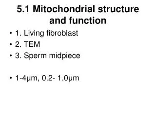
Mitochondrial DNA; responses to radiation, ageing & xenobiotics. David Maguire, PhD Griffith University, Nathan Campus, Brisbane, Australia Rochester, December 13 th , 2006. Mitochondrial anatomy; structure(s). Confocal microscopy, staining with Mitotracker dyes. human skin fibroblasts.

E N D
Mitochondrial DNA;responses to radiation, ageing & xenobiotics David Maguire, PhD Griffith University, Nathan Campus, Brisbane, Australia Rochester, December 13th, 2006
Mitochondrial anatomy; structure(s) Confocal microscopy, staining with Mitotracker dyes human skin fibroblasts bovine pulmonary artery endothelial cells Science Mar 5 1999 #5407 1483-1497 human placenta (E.M.) pancreatic cell
Outer membrane Inner membrane Cristae Mitochondria (orange) closely associate with microtubules of cytoskeleton
mitochondria evolution anatomy distribution functions citric acid cycle (tricarboxylic acid cycle) fatty acid oxidation electron transport chain (major ATP source) urea cycle (part) cellular calcium homeostasis plasma [NH3 ] control generation of ROS regulation of apoptosis) extranuclear inheritance mitochondrial genome homoplasmy/heteroplasmy segregation nuclear disorders of mitochondria urea cycle disorders of b-oxidation pathways electron transport chain disorders mitochondrial biogenesis disorders mtDNA disorders homoplasmic heteroplasmic threshold effect mtDNA & assays Enzymes Sequencing Deletion Depletion
Characterisitics (rat liver mitos) • Volume; 0.27 um3 • inner membrane area; 6.5um2 • complex I per mito; 2,600 - 15,600 • >100,000 electrons/sec [pyruvate as acceptor] • redox+ATPase complexes • ~7,500 per um2 • occupy ~ 40% of inner membrane • mean distance apart ~70-200 Angstroms • i.e. close enough to form micro-aggregates
Other mitochondrial function(s) • generation of reactive oxygen species (ROS) • superoxide (O2 .-), H2O2, OH. • Toxic byproducts of respiration • accumulate if early steps of ETC (I and CoQ) inhibited • appropriate ROS generated (destructive purposes) • macrophages - cell killing • inappropriate ROS generation • cell ‘suicide’
regulation of apoptosis (programmed cell death) • The primary step in the apoptosis cascade • opening of mt permeability transition pore (mtPTP) • permits release of • cytochrome C - acitivates the sytosolic caspase cascade • ‘apoptosis-inducing factor’ (AIF), a flavoprotein • translocates to nucleus and induces chromatin destruction • latent forms of caspases • caused by • excess Ca++ uptake by mt • increased exposure to ROS • decline in energetic capacity
ATP ATP food metabolism provides energy proteinscarbohydratesfats amino acidsglucoseglycerol + fatty acids glycolysis-oxidation pyruvate electrons + H+ acetylCoA NADH FADH2 citric acid cycleCO2electron transport chain • three major food sources
MITO - biochemistry 1 e- from NADH is passed down electron transport chain 2 one H+ pumped out of I, III & IV for each electron passing 3 H+ gradient created drives complex V rotation 4 complex V ‘rotation’ generates ATP (one molecule per H+ ) (3 per NADH) (2 per FADH2) H+ Inter memb space C ADP I III II IV V C1 a-Cu Q FeS ANT FeS a3-Cu memb Q FAD b matrix ADP +Pi ATP 1/2O2 H2O ATP H+ 2e- H+ NADH NAD+ succinate, FADH2 H+ H+
Subunits of ETC and ATP synthase • Most are nuclear-encoded • must be transported into mitochondria • Some vital ones mtDNA-encoded • (“extra-nuclear”, or “non-mendelian” inheritance) • efficient functioning requires • correct assembly of complexes • position • orientation • active sites
‘5kb common deletion’ region Human mitomap Swerdlow RH, Parks JK, Cassarino DS, Trimmer PA, Miller SW, Maguire DJ, Sheehan JP, Maguire RS, Pattee G, Juel VC, Phillips LH, Tuttle JB, Bennett JP Jr, Davis RE, Parker WD Jr. Mitochondria in sporadic amyotrophic lateral sclerosis.Exp Neurol. 1998 Sep;153(1):135-42. Swerdlow RH, Parks JK, Cassarino DS, Maguire DJ, Maguire RS, Bennett JP Jr, Davis RE, Parker WD Cybrids in Alzheimer's disease: a cellular model of the disease?Neurology. 1997 Oct;49(4):918-25. • 5kb deletion; • At site of 13 bp repeats • appears in • - ageing • late onset dementias • [See later]
5kb deLetion Genome dimerizes Part of genome deleted Leads to loss of a region of Mitochondrial DNA from one or more mtGENOMES
5 kb deLetion ASSAY Product made only in ‘long-time PCR Product made in ‘short-time PCR
human mt genome features • circular • contains ‘D-loop’ - includes ‘origin of replication’ • CODES FOR 37 MITOCHONDRIAL GENES • 2 rRNA genes (12S & 16S – ‘bacteria-like’ • 22 tRNA genes (one more than the theoretical minimum (20 amino acids) 2 for S & L • 13 subunits of mito. complexes • 7/39 NADH:ubiquinone oxidoreductase (I) • 1/10 ubiquinol:cytochrome C oxidoreductase(III) • 3/13 cytochrome C oxidase (IV) • 2/12 ATP synthase (V) • but 0 in succinate:UQ oxidoreductase or ANT
human mitochondrial genome very conservative of space (cf. yeast, etc.) • uses a non-universal code • lacks introns • many regions are read in both polarities • minimal tRNA genes • 22; (20 aminoacids so two redundant [S & L]) • stop codons truncated • have ‘exported’ to the nucleus many genes which remain mitochondrial in other species
further complication; heteroplasmy mitochondria heteroplasmic • homoplasmic wt homoplasmic mutant nucleus
heteroplasmic nucleus • homoplasmic heteroplasmichomoplasmic • wt mutant mutant SEGREGATION; allows somatic ‘drift’ and generation ‘gaps’
assess complexes I-V (enzyme assays) • time consuming • expensive • results are sometimes equivocal • appropriate source of mitochondria • [accessible tissue (leukocytes, platelets vs brain, cardiac muscle, skeletal muscle)] • uncertainty about contamination with non-mitochondrial material • cytoplasmic • other vacuoles • nuclear
sequencing the mitochondrial genome • big problem; heteroplasmy (polymorphism) • time consuming (sequence multiple clones/amplimers) • expensive (sequence multiple clones/amplimers) • results are often equivocal (number of clones?) • mitochondrial source (are all mitos equivalent?) • uncertainty about contamination with non-mitochondrial material • nuclear genes & pseudogenes (homology) • foreign genes • (technician, cell culture, bacterial, viral, etc)
Aminoglycosides induce deafness Some folk have a ‘more bacterial’ type of rRNA – more susceptible to these antibiotics Doxorubicin Inhibits mtDNA topoisomerase Inhibits mtDNA replication Inhibits mt DNA repair Inhibits mtDNA expression AZT Inhibits DNA polymerase Depletes mtDNA many others iatrogenic mitochondrial defects Other xenobiotics Unknown toxic material in jet fuel (Lintell, N, PhD thesis, 2006) Cause of up to 80% mtDNA depletion in a cohort of Australian airmen?
Mitochondrial population statistics Mitochondrial membrane mtDNA Nucleus Chromosomes (1-22, +XX) Most cells contain ~ 100 - 200 mitochondria (fertilized egg contains 100,000) Each mitochondria may contain 1 - 4 mt genomes (each 16,500bp)
dePletion Loss of one or more mtGENOMES Loss may be spread across Many mitochondria. Extreme case is rho0 cell, Which has no mtDNA left but Still has mitochondrial ‘ghosts’
mtDNA target Nuclear target = X mtDNA target Nuclear target < X dePletion ASSAY Two nuclear targets per cell 100 mt * 1 genomes = 100 mt targets per cell So, X = 100/2 = ~50 = ~ 4-5 PCR cycles
Human mitomap 46 bp tRNA [leucine] target (as per LIntell, 2006) [taqman] 526 bp ‘stable’ region target In humans (Murphy et al, 2006) [SYBR green} Murphy JE, Nugent S, Seymour C, Mothersill C. Mitochondrial DNA point mutations and a novel deletion induced by direct low-LET radiation and by medium from irradiated cells.Mutat Res. 2005 Aug 1;585(1-2):127-36.
Radiation effects • Murphy et al (2005) • In vitro 5mGy to 5Gy dose • mtDNA examined at times up to 96 hours • Increased deletion load • But not the common 5kb one! • Increased total mtDNA load • Conclusions; • Deletion; damage is occurring • Increased mtDNA; either • Smaller circles can replicate faster &/or uncontrolled? Or; • Damaged mtDNA not being removed? • (No-one’s emptying the trash?)
Our aims • Repeat Murphy’s work on irradiated cells • Deletion studies • Depletion studies • Develop taqman approach (quantitative) for depletion (& deletion) • Apply in vitro [mouse, Human] • Apply in vivo [mouse, Human, rat, dog…] • Apply to tissue ‘rescued’ from irradiation damage
M: 100 bp DNA ladder 1: mtDNA, 526 bp 2: Nuclear DNA, 155 bp Primers directed at human mtDNA and nuclear 18S RNA gene were used 3: Product from a pair of other primers 4: Human placenta total DNA only M1 2 3 4 mtDNA 526bp Nuc DNA 155bp Result 1 Dr Zhang Placental DNA Extracted & Amplified with Conventional PCR 2 tube depletion assay primers
Duplicate data mtDNA 18S RNA Result 2; real time 2 tube SYBR green Triplicate data mtDNA 18S RNA Threshold cycle (Tc) Melt curve of the two products Tc mit 1; 11.27 Tc mit 2; 12.79 Tc mit 3; 12.80 Tc nuc 1; 15.16 Tc nuc 1 ; 15.36 Tc nuc 1; 15.15 ~ 3 cycles apart (ergo threefold mt target excess)
DS DNA [minimum one copy] SS DNA 3’ primer 5’ primer&probe Primer extension & endonuclease activity of TAQ hydrolyses probe, releasing fluorophore and quencher DS DNA [2 * original copy number] Taqman 1 probe assay (better) 65’C 96’C 72’C 72’C 65’C Nett effect; concentration of free fluorophore doubles in each cycle
Two different fluorophores Two different probes Two different primer sets Taqman 2 probe assay Target 1 Target 2 65’C 96’C 72’C 72’C 65’C Nett effect; concentration of each free fluorophore doubles in each cycle Outcomes; ratio of one target compared to the other
Summary of orders for dePletion assay HUMANMOUSE
Summary of orders for deLetion assay HUMANMOUSE
‘5kb common deletion’ region The future?; 3 probe taqman design 1 46 bp tRNA [leucine] target (as per LIntell, 2006) [taqman] 2 3 Nuclear target 18SrDNA, or tubulin
conclusions • mtDNA damage is a feature of ageing • mtDNA is a known target for xenobiotics • Iatrogenic • Occupational • Both lead to depletion of mtDNA • mtDNA is also a target for radiationin vitro • Is it the major target for irradiation in vivo? • Therapies to protect mtDNA from damage? • IR damage to mtDNA seems different • Leads to increased mtDNA, rather than depletion!! • Up to 2 fold increase at 96 hours • Leads to increased burden of deleted mtDNA segments • Deletion appears different from ‘common 5kb deletion’ • 4881 cf 4997 bp long
Taqman assay designed for mouse mito assay using Biosearch Technologies software
Taqman probes designed vs mouse 18srDNA with FAM(biosearch software) • Function rank Tm GC% length [position] • Forward 87.63 59.1 53 17 [350-366] acgtctgccctatcaac • Reverse 74.76 58.6 56 16 [458-443] ggtagccgtttctcag • Probe 85.07 68.3 57 21 [367-387] FAMtttcgatggtagtcgccgtgcBHQ-1
Features of mouse mito taqman assay (calfluor red 635 & BHQ2)
Forward Primer (mito D-loop region) 5’ accgcggtcatacgattaac 3’ DMm1F(M) Reverse Primer (mito D-loop region)) 5’ cccagtttgggtcttagctg 3’ DMm1R(M) HYB OLIGO (MITO) 5’atcttcggcgtaaaacgtgt3’ DMm1Q(M)SEQUENCE SIZE: 660 Forward Primer (nuclear 18SrDNA) 5’ cgcggttctattttgttggt 3’ DMn1F(M) Reverse Primer (nuclear 18SrDNA) 5’ agtcggcatcgtttatggtc 3’ DMn1R(M) HYB OLIGO (NUCLEUS) 5’tgaggccatgattaagaggg3’ DMn1Q(M)SEQUENCE SIZE: 1869 [Same primers as two tube assay but with addition of sensor probe] Possible Mouse dePletion primers [one tube assay] designed by DM
Human depletion primers used by Nick [one tube assay] • Nuclear target; tubulin-a-8 • Forward sequence • 1759141 gcagccaacaactatgcccg 1759160 • Reverse sequence • `1759218 cttccgtatgcggtccagca 1759199 • Probe • 1759193 6-Fam-tcaatgctctccttgcccaccgtgtagt-BHQ1 1759166 • Mitochondrial target; tRNA leucine • Forward sequence • 3231gttaagatggcagagcccgg 3250 • Reverse sequence • 3299 gaagaggaattgaacctctgac 3278 • Probe • 3277 Rox-tgtaaagttttaagttttatgcgatta-BHQ23251
Mouse deLetion primers used[one tube, fast PCR assay] designed by DM • Forward (8300-8320) DMmdel1F (M) • 5’ ttgcccacttccttccacaag 3’ • Reverse (15962-15982) DMmdel1R (M) • 5’ gaaaggacagcacacagtcta 3’
Human deLetion primers used[one tube, fast PCR assay]as per Murphy • Forward DMmdF1 [H] • 5’ cgggggtatactacggtcaa 3’ • Reverse DMmdR1 [H] • 5’ ggtttcgatgatgtggtctttg 3’
Obvious addendum • mtDNA target in dePletion assay • Must not lie in a deletable region • Nor in a mutation hotspot region
NICK’S NEAT TRICKassay depletion AND deletion if use mito target in deletable regionDe(p)letion primers used[one tube assay with fluorometric dyes & FRET PCR]
Human de(p)letion primers used by Nick [one tube assay] • NUCLEAR Tubulin-a-8; amplified sequence size: 67 • Forward Primer [1759141 – 1759160] • 5’ gcagccaacaactatgcccg 3’ DMn1F(H) • Reverse Primer [1759218 – 1759199] • 5’ cttccgtatgcggtccagca 3’ DMn1R(H) • HYB OLIGO (nuc) [1759193 – 1759166] • 5 ’6-Fam-tcaatgctctccttgcccaccgtgtagt-BHQ1 ….3’ DMn1Q(H) • MITOCHONDRIAL tRNA leucine; amplified sequence size: 68 • Forward Primer [3231 – 3250] • 5’ gttaagatggcagagcccgg 3’ DMm1F(H) • Reverse Primer [3299 – 3278] • 5’ gaagaggaattgaacctctgac 3’ DMm1R(H) • HYB OLIGO (mito) [3277 – 3251] • 5’ Rox-tgtaaagttttaagttttatgcgatta-BHQ2 …..3’ DMm1Q (H)
Mouse dePletion primers used[one tube assay] designed by DM • Forward Primer (mito D-loop region) • 5’ accgcggtcatacgattaac 3’ DMm1F(M) • Reverse Primer (mito D-loop region)) • 5’ cccagtttgggtcttagctg 3’ DMm1R(M) • HYB OLIGO (MITO) 5’atcttcggcgtaaaacgtgt3’ DMm1Q(M)SEQUENCE SIZE: 660 • Forward Primer (nuclear 18SrDNA) • 5’ cgcggttctattttgttggt 3’ DMn1F(M) • Reverse Primer (nuclear 18SrDNA) • 5’ agtcggcatcgtttatggtc 3’ DMn1R(M) • HYB OLIGO (NUCLEUS) 5’tgaggccatgattaagaggg3’ DMn1Q(M)SEQUENCE SIZE: 1869 • [Same primers as two tube assay but with addition of sensor probe]
Overall plan • Develop & apply SYBR two tube assays to; • Mouse (normal & irradiated – various tissues) • Human (normal & irradiated – skin) • For both, tabulate • Radiation dose/response • Protective agents after radiation; dose/response • Apply deletion primer analysis to human • Apply taqman one tube, 2 site assays to; • Same as above (if positive above )
Ca2+ • Calcium plays a pivotal regulatory role in numerous cellular processes; therefore its cytosolic concentration undergoes very precise regulation. In electrically excitable cells calcium influx is controlled by the filling state of intracellular calcium stores. Depletion of them as well as Ca2+ flux into the cell are regulated by local Ca2+ concentration. Mitochondria may buffer Ca2+ in a close vicinity of Ca2+ channels and thereby influence both: the Ca2+ release from the stores and a feed back inhibition of Ca2+ channels in the plasma membrane. Dissipation of a mitochondrial membrane potential abolishes mitochondrial Ca2+ buffering capability what results in a deregulation of intracellular calcium signals.
MITO - biochemical genetics complexI III IIIVVANT mtDNA 710320 nDNA 329410101 total 3910413121 H+ C ADP IIIIII IVV C1 a-Cu Q FeS ANT FeS a3-Cu Q FAD b ADP +Pi ATP 1/2O2 H2O ATP H+ NADH succinate