Download
1 / 1

E N D
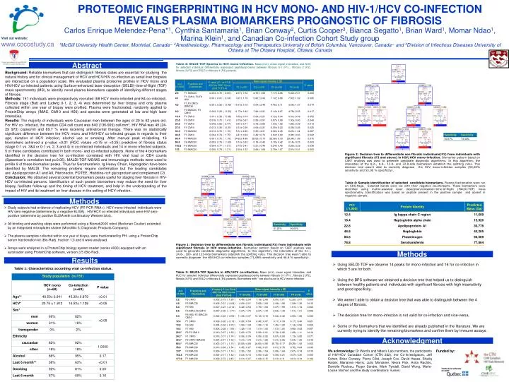
Visit out website: www.cocostudy.ca Table 2: SELDI-TOF Spectra in HCV mono-infection. Mass (m/z), mean signal intensities, and AUC for selected individual differentially expressed peptides/proteins between fibrosis 0-1 (F1) , fibrosis 2 (F2), fibrosis 3 (F3) and ESLD or fibrosis 4 (F4) patients. Abstract Background:Reliable biomarkers that can distinguish fibrosis states are essential for studying the natural history and for clinical management of HCV and HCV/HIV co-infection as serial liver biopsies are impractical on a population scale. We evaluated plasma proteome profiles in HCV mono and HIV/HCV co-infected patients using Surface-enhanced laser desorption (SELDI)-time-of-flight (TOF) mass spectrometry (MS), to identify novel plasma biomarkers capable of identifying different stages of fibrosis. Methods: 151 individuals were prospectively recruited (68 HCV mono-infected and 84 co-infected). Fibrosis stage (Batt and Ludwig 0-1, 2, 3, 4) was determined by liver biopsy and only plasma collected within one year of biopsy were profiled. Plasma were fractionated, randomly applied to ProteinChip arrays (IMAC, CM10 and H50) and spectra were generated at low and high laser intensities. Results: The majority of individuals were Caucasian men between the ages of 20 to 62 years old. For HIV co- infected, the median CD4 cell count was 640 (136-900) cell/mm3, HIV RNA was 49 (24-29 973) copies/ml and 89.7 % were receiving antiretroviral therapy. There was no statistically significant difference between the HCV mono and HIV/HCV co-infected groups in regards to their age, duration of HCV infection, alcohol use or smoking. After manual peak re-labeling, 16 biomarkers achieved a p-value <0.01 (ROC values >0.75 or <0.25) predictive of fibrosis status (stage 0-1 vs. 3&4 or 0-1 vs. 2, 3 and 4) in co-infected individuals and 14 in mono-infected subjects. 5 of these candidates contributed in both mono- and co-infected subjects. None of the 4 biomarkers identified in the decision tree for co-infection correlated with HIV viral load or CD4 counts (Spearman’s correlation test p>0.05). MALDI-TOF MS/MS and immunologic methods were used to profile 9 of these biomarker peaks. Thus far Serotransferin, Ig Heavy Chain, Haptoglobin have been identified by MALDI. The remaining proteins require confirmation but the leading candidates are: Apolipoprotein A1 and AII, Fibronectin, POTEE, Histidine-rich glycoprotein and complement C3. Conclusion: We obtained several potential biomarkers peaks useful for staging liver fibrosis in HIV-HCV co-infected persons. Identification of such protein biomarkers may reduce the need for liver biopsy, facilitate follow-up and the timing of HCV treatment, and help in the understanding of the impact of HIV and its treatment on liver disease in the setting of HCV infection. Proteomic Fingerprinting in HCV mono- and HIV-1/HCV Co-infection reveals plasma Biomarkers prognostic of fibrosisCarlos Enrique Melendez-Pena*1, Cynthia Santamaria1, Brian Conway2, Curtis Cooper3, Bianca Segatto1, Brian Ward1, Momar Ndao1, Marina Klein1, and Canadian Co-infection Cohort Study group1McGill University Health Center, Montréal, Canada~ 2Anesthesiology, Pharmacology and Therapeutics University of British Columbia, Vancouver, Canada~ and 3Division of Infectious Diseases University of Ottawa at The Ottawa Hospital, Ottawa, Canada Figure 2:Decision tree to differentiate non fibrotic individuals(F1) from individuals with significant fibrosis (F2 and above) in HIV/HCV mono-infection. Biomarker pattern based on CART analysis was used to generate candidate diagnostic algorithms. In this algorithm, the intensities of the 8,2-, 8.8-, 13.8- and 22.8-Kda biomarkers establish the splitting rules. This decision tree wasn’t able to correctly diagnose the HCV mono-infection samples (50,00% sensitivity and 55.00 % specificity). Table 4: Sample identification of selected candidate biomarkers. Plasma fractionation were run on SDS-Page. Selected bands were cut with their negative counterparts. These biomarkers were identified using matrix-assisted laser desorption/ionisation-time-of-flight (MALDI-TOF) mass spectrometry. Identification was based on peptide present in the positive sample and absent in negative sample. Methods • Study subjects had evidence of replicating HCV (RT-PCR RNA+). HCV mono-infected individuals were HIV sero-negative (determine by a negative ELISA). HIV/HCV co-infected individuals were HIV sero-positive (determine by positive ELISA with confirmatory Western blot). • All binding and washing steps were performed using a Biomek2000 robot (Beckman Coulter) extended by an integrated microplates shaker (MicroMix 5; Diagnostic Products Company). • The plasma samples collected within one year of biopsy, were fractionated by PH, using a ProteinChip serum fractionation kit (Bio-Rad), fraction 1,3 and 6 were analysed. • Arrays were analyzed in a ProteinChip biology system reader (series 4000) equipped with an autoloader using ProteinChip software, version 3.5 (Bio-Rad). Figure 1:Decision tree to differentiate non fibrotic individuals(F1) from individuals with significant fibrosis in HCV mono-infection. Biomarker pattern based on CART analysis was used to generate candidate diagnostic algorithms. In this algorithm, the intensities of the 4.1-, 24.8-, 100- and 133-Kda biomarkers establish the splitting rules. This decision tree wasn’t able to correctly diagnose the HIV/HCV co-infection samples (71,69% sensitivity and 46.6 % specificity). Methods Results • Using SELDI-TOF we observe 14 peaks for mono-infection and 16 for co-infection in which 5 are for both. • Using the BPS software we obtained a decision tree that helped us to distinguish between healthy patients and individuals with significant fibrosis with high insensitivity and good specificity.. • We weren’t able to obtain a decision tree that was able to distinguish between the 4 stages of fibrosis. • The decision tree for mono-infection is not valid for co-infection and vice-versa. • Some of the biomarkers that we identified are already published in the literature. We are currently trying to identify the remaining biomarkers and confirm them by immune assays. Table 1: Characteristics according viral co-infection status. Table 3: SELDI-TOF Spectra in HIV/HCV co-infection. Mass (m/z), mean signal intensities, and AUC for selected individual differentially expressed peptides/proteins between fibrosis 0-1 (F1) , fibrosis 2 (F2), fibrosis 3 (F3) and ESLD or fibrosis 4 (F4) patients. Biomarkers with * are also found in HCV mono-infection. Acknowledgment We acknowledge: Dr Ward’s and Ndao’s Lab members, the participants of HIV-HCV Canadian Cohort (CTN 222), the Co-Investigators, Jeff Cohen, Brian Conway, Pierre Côté, Joseph Cox, David Haase, Shariq Haider, Marianne Harris, Julio Montaner, Neora Pick, Anita Rachlis, Danielle Rouleau, Roger Sandre, Mark Tyndall, David Wong, Marie-Louise Vachon and the study coordinators/ nurses. Funded by:


