Download
1 / 24
240 likes | 243 Views
Craniovertebral Risk Factors Screening. Mobilization/Manipulation Cervical Spine. Ruling out the presence of cardinal signs and symptoms before mobilizing the C spine is a priority. Suggestive of either vertebral/basilar artery insufficiency, or cervical cord compression. Vertebral Artery.

E N D
Mobilization/Manipulation Cervical Spine • Ruling out the presence of cardinal signs and symptoms before mobilizing the C spine is a priority. • Suggestive of either vertebral/basilar artery insufficiency, or cervical cord compression.
Vertebral Artery • Arises from the first part of the subclavian artery and passes upward on the longuscolli to enter the foramen transversarium of C6 • It then ascends from C6 to C1. After emerging through the transverse foramen of C1, it winds around the articular pillar and together with the 1st cervical nerve and veins pierces the posterior atlanto-occipital membrane to enter the cranium through the foramen magnum • On the anterior side of the brainstem it joins its fellow to form the basilar artery • The vertebral arteries contribute about 11 percent of the total cerebral blood flow, the remaining 89 percent being supplied by the carotid system.
Ligaments of C1-2 Cruciate ligament • The major portion of this ligament is the transverse ligament • There is an ascending and a descending part, which are triangular shaped • Check inferior/superior displacement of the transverse ligament. Transverse ligament • Most important ligament in upper cervical spine • 7-8 mm thick. • Attaches on the medial surface of the lateral mass of the atlas
Signs/Symptoms of Cord Compression • Bilateral or quadrilateral limb paresthesiae • Hyperreflexia • Clonus • Positive Babinski or Hoffman’s • Arm and leg weakness • Lack of coordination bilaterally
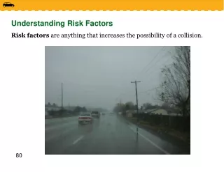
Risk factors Cervical Arterial Dysfunction • History of trauma to cervical spine / cervical vessels • History of migraine-type headache • Hypertension • High cholesterol levels • Cardiac disease, vascular disease, previous cerebrovascular accident or transient ischaemic attack • Diabetes mellitus • Blood clotting disorders
Risk factors Cervical Arterial Dysfunction • Anticoagulant therapy • Long-term use of steroids • History of smoking • Recent infection • Immediately post partum • Trivial head or neck trauma • Absence of a plausible mechanical explanation for the patient’s symptoms.
Differential Diagnosis • Vertebral artery insufficiency • Alar ligament insufficiency • Transverse ligament insufficiency • C 1 - 2 instability • Jefferson fracture • Balance difficulty, due to loss of proprioception secondary to immobilization of cervical spine • Symptom magnification • Autonomic reactions
Presentation • Drop attacks • Dizziness • Dysphagia (difficulty swallowing) • Dysarthria (speech change, either slurred or slowed) • Diplopia (double vision) • Nausea • Nystagmus • Facial numbness/lip paresthesia
Upper Cervical Stability Testing • Transverse Ligament • Alar Ligament • Jefferson fracture
Pre-screening Vertebral Artery • Current research does not support the contention that provocative positional testing can accurately identify patients at risk for cervical artery disease • Vertebral artery testing procedures have a sensitivity and specificity that approximates zero: high likelihood of false negative findings. • Test procedures for the vertebral artery also hold a certain risk, and screening tests will not identify all patients at risk of suffering adverse reaction to cervical manipulation
Observation and History • This is an opportunity to observe and recognize possible red flag indicators such as gait disturbances, subtle signs of balance problems, upper motor neuron signs, cranial nerve dysfunction, and behavior suggestive of upper cervical instability (e.g. anxiety, supporting head/neck) early in the clinical encounter.
How to proceed in the absence of certainty • Cervical artery strokes are rare, but are an important consideration as part of your assessment • Arterial dissection and other vascular presentations are fairly recognizable if the appropriate questions are asked during the patient history, • Based on the current evidence, there is no strong foundation for the claim that there is a causal relationship between cervical manipulation and vertebral artery dissection or stroke. • There is no strong empirical evidence to support the notion that upper cervical manipulation carries any greater risk of injury than middle or lower cervical manipulation, or that non thrust mobilization to any region of the cervical spine carries any less risk than manipulation to the same region
How to proceed in the face of uncertainty • Upper cervical manipulations, especially the ones involving rotation, should be used with caution. • Stay away from endrange techniques, especially extension and rotation • Develop a high index of suspicion for cervical vascular pathology, especially with cervical trauma
How to proceed in the absence of certainty • Consider limitations of current objective tests. • In cases of acute onset headache “unlike any other”, conservative treatment techniques are recommended in the early stages • Do not further examine or treat a patient with signs and symptoms of cervical artery disease
How to proceed in the absence of certainty • You cannot ignore the presence of cardinal signs and symptoms • A patient with cardinal signs and symptoms warrants a referral to a medical specialist for appropriate management • Incorporate T spine manipulation in the treatment of C spine disorders.
Alternative Approach to Direct Cervical Treatment • Current evidence suggests a large likelihood of improved patient outcomes when thoracic manipulation is coupled with cervical active range of movement exercises • Subsequent sessions can then introduce more direct manual cervical treatments if warranted
VBI and Upper Cervical Stability Screening Protocol • Expert history taking • Upper cervical ligamentous stability testing • Sustained end range cervical rotation to the left and the right. Ask about dizziness during each test/ Observe the eyes for the presence of nystagmus • The position or movement that provokes symptoms as described by the patient • Sustained mobilization position • Always assess prior and immediately after a technique involving endrange rotation, as well as first thing next visit…