Download
1 / 37
450 likes | 540 Views
Molecular Biology Techniques. Part 1. Learning Objectives. This lecture reviews some of the techniques used in molecular biology; the methods that concern themselves with analysis of the structure & function of DNA & RNA at the molecular level. These techniques are important in:.

E N D
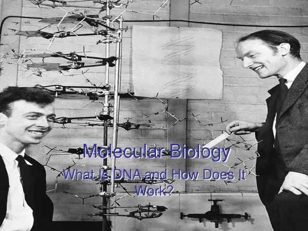
Molecular Biology Techniques Part 1
Learning Objectives • This lecture reviews some of the techniques used in molecular biology; the methods that concern themselves with analysis of the structure & function of DNA & RNA at the molecular level. • These techniques are important in: • Diagnosis of genetic disorders (e.g. cystic fibrosis). • Diagnosis of certain infective disorders (e.g. human immunodeficiency virus (HIV) and hepatitis B & C infections). • Investigation of the molecular basis of cancer. • Studying common polygenic disorders (e.g. essential hypertension and diabetes mellitus).
Polymerase Chain Reaction (PCR) Invented in 1993 by Kary Mullis Received a Nobel Prize in chemistry in 1993, for his invention of the polymerase chain reaction (PCR). Application:in vitro amplification of DNA segments, without using a living organism (such as E. coli or yeast).
PCR Revolutionized the Study of Genes • PCR allows a small section of human genomic DNA to be amplified many times in the test tube. • The technique is simple, fast, and requires as little as a single cell. • DNA for analysis may be obtained from any nucleated cell; most commonly used are WBCs, but hair roots, mouth scrapings, or sperm can also be used. • Great care must be taken to avoid contamination with cells or DNA from the laboratory. • Most PCR methods typically amplify DNA fragments of up to 10 kilo base pairs (kb), although some techniques allow for amplification of up to 40 kb.
PCR in Practice • Basic components are: • DNA template that contains the region of the DNA fragment to be amplified • Primers, which are complementary to the DNA regions at the 5' and 3' ends of the DNA region that is to be amplified. • DNA polymerase (e.g. Taq polymerase or another DNA polymerase with a temperature optimum at around 70°C), used to synthesize a DNA copy of the region to be amplified • Deoxynucleotide triphosphates, (dNTPs) from which the DNA polymerase builds the new DNA • Buffer solution, which provides a suitable chemical environment for optimum activity and stability of the DNA polymerase • Divalent cation, magnesium or manganese ions; generally Mg2+ is used • Monovalent cation potassium ions The PCR is carried out in small reaction tubes (0.2-0.5 ml volumes), containing a reaction volume typically of 15-100 μl, that are inserted into a thermal cycler. This is a machine that heats and cools the reaction tubes within it to the precise temperature required for each step of the reaction. The PCR usually consists of a series of 20 to 35 cycles.
PCR ( Polymerase chain reaction)How to magnify a desired gene?
How to Express Results ? To check whether the PCR generated the anticipated DNA fragment, agarose gel electrophoresis is commonly employed for size separation of the PCR products. The size(s) of PCR products is thereby determined by comparison with a DNA ladder, which contains DNA fragments of known size, ran on the gel alongside the PCR products Ethidium bromide-stained PCR products after Gel electrophoresis. 2 sets of primers were used to amplify the IGF gene from 3 different DNA samples. In sample #1 the gene was not amplified by PCR, bands for tissue #2 and #3 indicate successful amplification of the IGF gene. A +ve control, and a DNA ladder containing DNA fragments of defined length (last lane to the right) to estimate fragment sizes in the experimental PCRs, were also ran on this gel.
Practical modifications to PCR technique Nested PCR - increases the specificity of DNA amplification, by reducing background due to non-specific amplification of DNA. Two sets of primers are being used in two successive PCR reactions. (Reverse Transcription PCR) RT-PCR - a method used to amplify, isolate or identify a known sequence from a cellular or tissue RNA. PCR reaction is preceded by a reaction using reverse transcriptase to convert RNA to cDNA. RT-PCR is widely used in expression profiling, to determine the expression of a gene or to identify the sequence of an RNA transcript. Quantitative PCR - used to measure the quantity of a PCR product . It is the method of choice to quantitatively measure starting amounts of DNA, cDNA or RNA. Real Time PCR – developed because of: 1) The need to quantitate differences in mRNA expression & 2) The availability of only small amounts of mRNA. Real Time PCR is a kinetic approach in which you look at the reaction in the early stages while it is still linear . SYBR Green, which has very low fluorescence in the absence of double stranded DNA and very high fluorescence in the presence of double stranded DNA is commonly used.
Clinical Applications of PCR • Allow the amplification & analysis of the DNA in a single cell, hair follicle, sperm, … to serve investigations in forensic medicine. • Detection of infectious agents, especially latent viruses. • Prenatal genetic diagnosis of mutations. • Detection of allelic polymorphism. • Establishing precise tissue types for transplants. • Study of evolution, using DNA from archaeological sample.
Blotting Methods • Southern blotting involves the transfer of DNA from a gel to a membrane, followed by detection of specific sequences by hybridization with a labeled probe. • Northern blotting, RNA is run on a gel. • Western blotting entails separation of proteins on an SDS gel, transfer to a nitrocellulose membrane, and detection proteins of interest using antibodies.
Southern Blotting Named after Dr. Edward M. Southern who developed this procedure at Edinburgh University in the 1970s Application: Allows detection of DNA fragments by hybridisation to a suitable probe.
Procedure: • DNA is cut first with restriction endonucleases. • The cleaved DNA fragments are separated on the basis of size using agarose gel electrophoresis. • After alkaline denaturation, the DNA fragments are transferred to a nitrocellulose membrane (the blot), by layering the nitrocellulose membrane over the gel (which rests in buffer). The gel is placed on adsorbent paper which acts as a wick to draw buffer through the gel and enhance DNA transfer to the membrane. The membrane is much easier to handle than the fragile agarose. • The DNA can be analysed with labelled probes that will hybridise to complementary sequences.
G A T C A G T A G Finding your gene of interest • DNA hybridization • find sequence of DNA using a labeledprobe • short, single stranded DNA molecule • complementary to part of gene of interest • labeled with radioactive P32 or fluorescent dye • heat treat DNA in gel • unwinds (denatures) strands • wash gel with probe • probe hybridizes with denatured DNA labeled probe genomic DNA C T A G T C A T C
Southern blotting blot DNA off of gelonto filter paper restriction digest gel electrophoresis expose filter paper toX-ray film wash filter with labeled probe
Southern blotting Edwin Southern Southern blotIDing one gene Southern blotillustration gel of genomic DNA
Clinical Applications of Southern Blotting • Detection of presence & determination of the number of copies of particular sequence or gene in genomic DNA, e.g. determine if liver has a gene for insulin? or how many copies of LDL receptor gene are present in the liver of heterozygote FH patient as compared to normal individual? • Diagnosis of single-gene defects. e.g. in sickle-cell anaemia, the single a.a substitution of valine for glutamic acid is the result of a single-base change in which thymine replaces adenine. This single base change leads to the loss of the recognition sequence for restriction endonuclease termed MstIII in the mutant gene. When the genomic DNA is cut with this enzyme and subjected to southern blotting using a probe specific for the b-globin gene, the length of DNA containing the complementary sequence differs between the sickle cell and normal specimens. Similar to Southern blot, Northern (RNA) and Western (protein) blots are used to size and quantitate specific RNA and protein molecules, respectively.
Northern blotting is similar to Southern blotting, but involves the transfer of RNA from a gel to a membrane RNA
How to separate mRNA from all other classes of RNA mRNA contains ~200 oligo(A) residues at 3’ end FIGURE 22: Poly(A)+ RNA can be separated from other RNAs by fractionation on an oligo(dT) column
Northern blotting: Measuring gene activity Poly(A)+ RNA: from rat tissues Probe: G3PDH (glyceraldehyde-3-phosphate dehydrogenase) From Dr. Yu
Western blotting • Western blotting entails separation of proteins on an SDS gel, transfer to a nitrocellulose membrane, and detection proteins of interest using antibodies. wikipedia
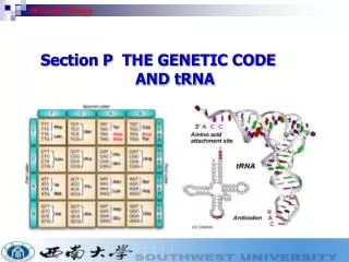
MOLECULAR BIOLOGY – Molecular biology techniques NUCLEIC ACID HYBRIDIZATION labeled probe Fluorescence In Situ Hybridization (FISH)
MOLECULAR BIOLOGY – Molecular biology techniques 32P Y anti-DIG antibody Probe labeling by incorporation of modified (d)NTPs radioactive labeling AUTORADIOGRAPHY Fluorophores conjugation Enzymes alkaline phosphatase horseradish peroxidase Streptavidin Biotin-11-dUTP (DIG) chemiluminiscence
MOLECULAR BIOLOGY – Molecular biology techniques DIG DIG DIG DIG DIG DIG DIG DIG DIG DIG DIG HRP HRP HRP HRP HRP HRP light light light light light light Y Y Y Y Y Y substrate substrate substrate substrate substrate substrate
MOLECULAR BIOLOGY – Molecular biology techniques GC rich Temp Temp AT rich GC rich DNAdenaturation Melting (denaturation) temperature depends on these major factors: - GC content (and therefore AT content) - sequence length - gaps in the annealed strands - salt concentration - pH - organic solvents (DMSO, formamide...)
MOLECULAR BIOLOGY – Molecular biology techniques CHROMOSOME PAINTING – MULTI COLOR FISH
Detection of Hepatitis C virus (HCV) infection • Based on the technique of hybridization: • HCV is a single-stranded RNA virus in the Flaviviridae family. The genome is approximately 10,000 nucleotides and encodes a single polyprotein of about 3,000 amino acids. • If an individual is infected with a virus (e.g. HCV), the genetic material of the virus will be different from the individual’s DNA or RNA. • RNA (including viral RNA) is isolated from patient’s blood. • Detect viral RNA by RT-PCR using primers targeting the HCV sequence.
MOLECULAR BIOLOGY – Molecular biology techniques How to see genes ... How to see specific forms of genes? How to clone and amplify genes? ... where in tissues? ... where on chromosomes? in situ hybridization recombinant DNA southern blot genomic libraries RFLP
References: • Clinical Biochemistry: An illustrated colour text, A. Gaw et al., Churchill Livingstone. • Clinical Chemistry, W.J. Marshall, Mosby. • Enzyme Tests in Diagnosis, D.W. Moss & S.B. Rosalki, Arnold. • Clinical Chemistry: Interpretation and Techniques, A. Kaplan & L.L. Szabo, Lea & Febiger, Philadelphia. • Fundamentals of Clinical Chemistry, N.W. Tietz, W.B., Saunders Company, Philadelphia, London, Tokyo …. • Clinical Biochemistry, A.F. Smith, G.J. Beckett, S.W. Walker & P.W. H Rae, Blackwell Science Ltd, London… • Interpretation of Diagnostic Tests, I. Wallach, Little, brown & Company, Boston, London ….. • Practical Clinical Biochemistry, H. Varley, A.H. Gowenlock & M. Bell, William Heinemann Medical Books LTD, London