Download
1 / 20
200 likes | 236 Views
Case Study: A patient with a history of alcoholism and bilateral pneumonia. Patient presents to the emergency department. A 59-year-old male patient presents to the ED complaining of acute chest pain and productive cough for 24 hours

E N D
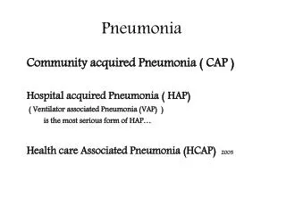
Case Study:A patient with a history of alcoholism and bilateral pneumonia
Patient presents to the emergency department • A 59-year-old male patient presents to the ED complaining of acute chest pain and productive cough for 24 hours • At presentation, he has moderate hypothermia (36.0°C), tachypnoea, tachycardia, SaO2 87% with FiO2 0.4 and BP 80/60 mmHg • Laboratory tests reveal: • Leukopenia (1100/mm3, mainly polymorphonuclear leukocytes [PMNs]) • Anaemia (haematocrit 26.5%, haemoglobin 9.1 g/dL) • High erythrocyte sedimentation rate (ESR) (115 mm/first hour) • Chest X-rays show extensive infiltrations in the right superior and left medium lung zones
History • The patient’s history reveals hospitalisation 2 years ago due to recurrent episodes of coughing and small volume hemoptysis with weight loss • He is a mariner (last sailed 6 months ago) and a habitual alcohol drinker • A chest CT scan from the previous hospitalisation demonstrates the existence of three cavitational lesions in the right lung
Laboratory findings and treatment2 years ago • Persistent laboratory investigations fail to confirm a diagnosis of TB • Laboratory investigations for vasculitis and other auto-immune diseases are also negative • Despite this, he receives empiric anti-TB treatment for 1 month without clinical improvement, but there is radiological deterioration • Sputum cultures are not diagnostic and blood cultures are negative • Under the presumed diagnosis of aspiration pneumonia, he receives antimicrobial therapy with ciprofloxacin plus clindamycin, after which he has substantial clinical improvement • He is discharged from hospital, but does not return for the recommended follow-up
Patient in ED: Questions • What is your differential diagnosis? • What is your opinion on the antimicrobial therapy? Which of the following would you include in a combination regimen? • Fusidic acid • Azithromycin • Fluconazole • Ampicillin/sulbactam • Piperacillin/combactam • A carbapenem
Question • What is your recommendation for the patient’s therapy? Consider options other than the antimicrobial therapy
Clinical course in the ED • An empiric combination therapy is commenced with: • Fusidic acid • Azithromycin • Fluconazole • Ampicillin/sulbactam • During the current hospitalisation, the patient’s condition worsens within hours, necessitating admission to the ICU. He is: • Mechanically ventilated • Transfused 1 unit of red blood cells • Microbiological investigations are performed on blood and bronchial secretions
Further clinical course • He develops galactic (lactic) acidosis, hemodynamic instability, and he spikes a high fever (>39.5°C) and leukocytosis (which gradually reaches 23000/mm3with a predominance of PMNs) • But then his condition improves and four days later, he is discharged from the ICU as he is: • Afebrile, hemodynamically stable with normal blood gases,but with a very slight regression of his leukocytosis (20000/mm3)
Further clinical course • The next day, his fever recurs • Klebsiella pneumoniae is isolated from both blood cultures and bronchial secretions (which had been sent from the ICU) and the tip of the central vein catheter reveals presence of Acinetobacter baumannii
Results of sensitivity testing *K. pneumoniae is found to produce extended-spectrum β-lactamase (ESBL) and metallo-β-lactamase (MBL)
Questions • The patient is again febrile. What do you think is the reason for this? e.g. consider whether treatment had failed, superinfection etc • Which antimicrobial treatment would you recommend now?
Patient is transferred • Due to multiresistant infection, the patient is transferred to the Infection Unit of a tertiary care hospital • Upon admission, he is: • Febrile (38.8°C), hypoxaemic (PO2 55mmHg), with leukocytosis (16200/mm3)but without marked PMN (PMN 78%), ESR 105 mm/first hour, procalcitonin (PCT)0.17 ng/mL • Seriously malnourished and suffering from alcoholic polyneuritis, resulting in an inability to stand or walk
Clinical course • A new sputum culture is obtained • He receives combination antibiotic therapy with meropenem 1g x 3 IV andcolimycin2 MU x3 IVand 1 MU x 3 by inhalation • Three days later, his fever is slightly reduced (~38°C), leukocytes have decreased to 12700/mm3 and he is hemodynamically stable • A chest CT scan reveals bilateral cavitations
Test results • Amplified Mycobacterium tuberculosis direct test (Gen-Probe) and Ziehl-Neelsen smear are negative • Non-quantitative sputum culture reveals presence of MRSA • Question: What is your suggestion now?
Empiric therapy remains the same • The empiric regimen is not modified • Three days later, the patient is again afebrile with normal blood gases and a normal leukocyte count • During the following days, he has gradual radiological improvement • Nevertheless, he remains unable to rise from his bed; he has a coarse tremor and very severe muscle atrophy • The total duration of antimicrobial therapy is 15 days • Two days later, he is transferred to a rehabilitation centre and re-evaluation is requested after 3 months
Questions • Do you agree with the duration of the treatment? • What general comments would you like to make regarding this case? • How might your clinical practice be influenced as a result of this case?
Comments • The infection was most likely due to the K. pneumoniae (in blood and respiratory secretions) • The organism probably developed resistance to the empiric therapy • The MRSA was likely to be a colonising organism • No antibiotic therapy was given specifically to treat infection with MRSA