Download
1 / 26
260 likes | 494 Views
Three-Dimensional Human Airway Segmentation for Sleep Apnea Diagnosis using Tubular Deformable Organisms. Tricia Pang November 25, 2008. OVERVIEW. Motivation Approach Preliminary Investigation Deformable Organisms Preliminary Results Conclusion. OVERVIEW. Motivation Approach

E N D
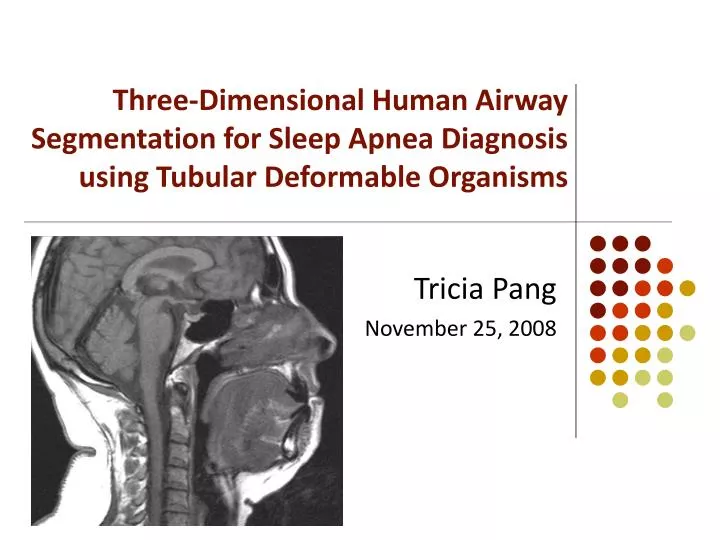
Three-Dimensional Human Airway Segmentation for Sleep Apnea Diagnosisusing Tubular Deformable Organisms Tricia Pang November 25, 2008
OVERVIEW • Motivation • Approach • Preliminary Investigation • Deformable Organisms • Preliminary Results • Conclusion
OVERVIEW • Motivation • Approach • Preliminary Investigation • Deformable Organisms • Preliminary Results • Conclusion
Motivation • Obstructed sleep apnea (OSA) disorder • Caused by collapse of soft tissue walls in the airway → model patient's airway to help diagnosis • Hand-segmentation: laborious • Goal: to develop automated tool for creating a patient-specific model of the airway Credit: Wikipedia
Motivation • Artisynth [2] &OPAL Project(OPAL = Dynamic Modeling of the Oral, Pharyngeal and Laryngeal Complex for Biomedical Engineering) • Import resulting airway into dynamic throat and mouth model for simulation
OVERVIEW • Motivation • Approach • Preliminary Investigation • Deformable Organisms • Preliminary Results • Conclusion
Data Source - MRI • Normal subjects, OSA patients, various treatments • Volumetric and cross-sectional measurements
OVERVIEW • Motivation • Approach • Preliminary Investigation • Deformable Organisms • Preliminary Results • Conclusion
Preliminary Investigation • Combined 2D segmentation of axial slices in Matlab • Procedure: • User-indicated start point at base of airway • Starting on axial slice at start point, grow ellipse outward • Iterate on all axial slices moving upwards along airway, and use previous segmentation as starting contour • “Active contours without edges” (Chan-Vese) [1]: • Based on Mumford-Shah framework • Evolve curve by minimizing energy from image (interior/exterior mean) and curvature
OVERVIEW • Motivation • Approach • Preliminary Investigation • Deformable Organisms • Preliminary Results • Conclusion
Deformable Organism • I-DO: framework for ITK (McIntosh & Hamarneh) [4] • Geometrical and physical layers of classical deformable models (data-driven) • Behavioral and cognitive layers for intelligent deformation control (knowledge-driven) • Related work: • Spinal crawler [5] • Vessel crawler [6]
Deformable Organism • Goal: automatically segment airway by growing a tubular organism, guided by image data and a priori anatomical knowledge • Advantages: • Increased accuracy • Analysis and labeling capabilities • Ability to incorporate shape-basedprior knowledge • Modular framework
Summary of Layers Control Center Grow, terminate, (branch) Sensors ‘GrowSense’ ‘HessianSense’ (‘BranchSense’) Behavior Grow, fit, (branch) Physics/Deformation Spring-mass system Medial and boundary nodes Radial, circumferential and sheer springs Geometric Medial-based shape representation Tubular with symmetric cross-section (often elliptical)
Viewer Adaptor • Graphical interface for viewing geometry of DOs and their deformations in real time
OVERVIEW • Motivation • Approach • Preliminary Investigation • Deformable Organisms • Preliminary Results • Conclusion
OVERVIEW • Motivation • Approach • Preliminary Investigation • Deformable Organisms • Preliminary Results • Conclusion
Summary • Model of a patient’s airway valuable to diagnosing the OSA disorder • Tubular deformable organisms • spring-mass system initiated at a user-indicated point • grown along the airway boundary using a priori knowledge of upper airway anatomy
References [1] Chan, T. and Vese L. Active Contours Without Edges. IEEE Transactions on Image Processing, 10 (2001) [2] Fels, S., et al. Artisynth: A biomechanical simulation platform for the vocal tract and upper airway. International Seminar on Speech Production (2006) [3] Hamarneh, G. and McIntosh, C. Physics-Based Deformable Organisms for Medical Image Analysis. Proc of SPIE 5747 (2005) 326-335 [4] McIntosh, C. and Hamarneh, G. I-DO: A “Deformable Organisms” framework for ITK. Medical Image Analysis Lab, SFU. Release 0.50. [5] McIntosh, C. and Hamarneh, G. Spinal Crawlers: Deformable Organisms for Spinal Cord Segmentation and Analysis. MICCAI (2006) 808–815 [6] McIntosh, C. and Hamarneh, G. Vessel Crawlers: 3D Physically-based Deformable Organisms for Vasculature Segmentation and Analysis. Proceedings of IEEE CVPR (2006)