Download
1 / 117
1.17k likes | 1.27k Views
SECTION II ADULT HEALTH NURSING. UNIT 12 NURSING CARE OF THE CLIENT: MOBILITY, COORDINATION, AND REGULATION CHAPTER 39. SENSORY SYSTEM (Eye) Part 2 Leslie Lehmkuhl, RN 2009. The eye is the organ of sight, a nearly spherical hollow globe filled with fluids (humors).

E N D
SECTION II ADULT HEALTH NURSING UNIT 12NURSING CARE OF THE CLIENT: MOBILITY, COORDINATION, AND REGULATION CHAPTER 39 SENSORY SYSTEM (Eye) Part 2 Leslie Lehmkuhl, RN 2009
The eye is the organ of sight, a nearly spherical hollow globe filled with fluids (humors). The outer layer or tunic (sclera, or white, and cornea) is fibrous and protective. The middle tunic layer (choroid, ciliary body and the iris) is vascular. The innermost layer (the retina) is nervous or sensory. The fluids in the eye are divided by the lens into the vitreous humor (behind the lens) and the aqueous humor (in front of the lens). The lens itself is flexible and suspended by ligaments which allow it to change shape to focus light on the retina, which is composed of sensory neurons Eye Anatomy
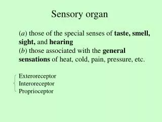
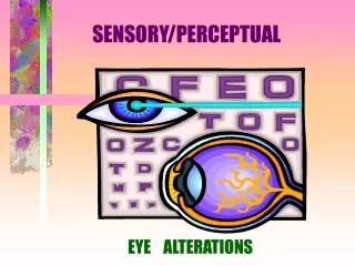
The Eye Accessory structures: eyebrows, eyelashes, eyelids, lacrimal apparatus, conjunctiva Lacrimal apparatus- manufacture and drains tears, contains lysozyme a bactericidal enzyme Conjunctiva- membrane that lines inner aspect of eyelids and anterior surface of the eyeball Anatomy of the eye 3 layers: sclera, choroid, and retina Sclera- outermost layer of the eye, made of white connective tissue cornea Choroid- dark brown membrane that lines internal area of the sclera, vascular layer pupil Retina- nervous tissue membrane that receives images and transmits impulses to the optic nerve Cones, rods, optic disk Sensory Disorder
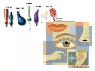
The eye is divided into two chambers by the crystalline lens Anterior chamber- contains aqueous humor Watery fluid Maintains normal intraocular pressure Posterior chamber- contains vitreous humor Jelly like Gives shape to the eyeball Physiology of vision Light must travel Through the cornea Through the aqueous humor Through the pupil Through the lens Through the vitreous humor To the cones and rods of the retina Processes necessary to form an image: refraction, accommodation, constriction, convergence Sensory Disorder
Diagnostic eye examinations Snellen test- screening test which assesses visual acuity Patients stands 20 feet away covering one eye, and then the other 20/20 normal vision Color vision charts Amster grid Refraction- determines refractory errors using retinoscope or lenses Refractory errors Astigmatism- defect in curvature of the eyeball surface causing blurred vision Strabismus- cross eyed vision due to asymmetry Myopia- inability to see distance (nearsighted) Hyperopia- inability to see close (farsighted) Presbyopia- changes in accommodation due to aging (usually in 40s) Sensory Disorder
Vision • Retinal image
Refraction: light rays are bent as they pass through colorless structures in eye –allowing light to focus on retina Accommodation: eye can focus on objects at different distances image on retina is changed by curvature of lens Vocabulary
Constriction:size of pupil controlled by dilator and constrictor muscles of iris Convergence: convergence medial movement of both eyes allows light rays to hit same point on retinas Vocabulary
Snellen’s test • Visual acuity • Client 20 feet away from chart • Covers one eye • Reads line above or below 20/20 • Line repeats with other eye
Visual acuity • Snellen chart • Checks vision at 20 feet from chart 20/20 • PERRL pupils equal, round, react to light • PERRLA pupils, equal, round, react to light, accommodate
Visual acuity tests may be performed in many different ways. It is a quick way to detect vision problems and is frequently used in schools or for mass screening. Driver license bureaus often use a small device that can test the eyes both together and individually. Snellen Chart Visual acuity test
slit-lamp, which is a specialized magnifying microscope, is used to examine the structures of the eye (including the cornea, iris, vitreous, and retina). The slit-lamp is used to examine, treat (with a laser), and photograph (with a camera) the eye. Slit-lamp exam
Disorder that causes the lens or its capsule to lose its transparency and/or become opaque.. Noninfectious opacity or clouding of the lens Congenital, exposure to systemic disease, trauma, toxins, medications as steroids, aging (most common cause).. As clouding develops, visual impairment occurs S/S: blurred vision, diplopia, photosensitivity, difficulty driving at night, glare, opacity in center portion of lens, hazy vision, glare, difficulty driving at night. The only treatment for a cataract is surgical removal of the lens Cataract extraction Intracapsular- not common Extracapsular- removes lens and anterior capsule with intracapsular lens implant CATARACTS
This photograph shows a cloudy white lens (cataract) over the pupil. Cataracts are a leading cause of decreased vision in older adults, but children may have congenital cataracts. With surgery, the cataract can be removed, a new lens implanted, and the person can usually return home the same day. Cataract - close-up of the eye
Cataract surgery - series: Normal anatomy The lens of an eye is normally clear. A cataract is when the lens becomes cloudy as you get older Cataract Surgery: Normal
Surgery is usually recommended for people who have vision problems or other major problems caused by the cataract. Indications; Cataract Surgery
Two procedures are used to treat cataracts. In the manual extraction procedure, a small incision is made at the edge of the outer lining of the eye (cornea). The lens is then removed and replaced with an artificial lens. Cataract surgery - series: Procedure, part 1
Another procedure is called phacoemulsification. This involves inserting a needle through a small incision on the eye. The end of the needle produces sound waves. The sound waves break up the lens, which is then sucked out through the needle. This procedure requires a smaller incision than the manual extraction procedure. Cataract surgery - series: Procedure, part 2
Cataract surgery usually works very well. The operation has few risks, the pain and recovery period are short, and your sight is usually greatly improved. Ninety-five percent or more of all cataract surgeries result in improved vision. Initially will have difficulty with images and depth perception Teach to avoid activities that could cause potential injury.. No coughing, sneezing, dark glasses to decrease glare, wear eye patch when sleeping. Cataract Surgery-Series: Aftercare
Post operative: no coughing , sneezing, vomiting, bending No increase in intraocular pressure after surgery Sleep on unoperative side Eye patch on at night for several nights Eye surgery
Disorder characterized by an elevated high pressure of fluid inside the eyeball d/t obstruction of outflow of aqueous humor. Treatment is focused on drug therapy to reduce intraocular pressure. Surgical intervention to facilitate drainage of aqueous humor is called an iridectomy. GLAUCOMA
Glaucoma is a condition of increased fluid pressure inside the eye. The increased pressure causes compression of the retina and the optic nerve which can eventually lead to nerve damage. Glaucoma can cause partial vision loss, with blindness as a possible eventual outcome. Glaucoma
Elevated pressure within the eye due to obstruction of outflow of aqueous humor Open angle: no signs or symptoms in early stage… then tunnel vision, eye pain , diffficulty adjusting to darkness, halo around lights, inability to detect colors Glacuoma
Glacuoma • Closed angle: severe pain, decreased vision, nausea, vomiting, sclera erythematous, and pupil enlarged and fixed colored halos around lights • Increased intraocular pressure • Open angle—beta blockers, miotics • Closed angle—osmotic diuretics, miotics, surgery….
RETINAL DETACHMENT • Separation of the retina from the choroid. • This condition is painless and may result from trauma or from intraocular disorders. • Treatment includes procedures that create an inflammatory reaction that results in retina reattaching to the choroid, or surgery.
Retinal detachment • Cryosurgery to freeze borders of retinal hole • Diathermy burn retina for sealing scarring occurs • Cryosurger to freeze border4s of retina hole • Scleral buckling • Eye patch for 1-2 days after surgery patch both eyes
Retina • Diabetic retinopathy • Microaneurysms, hemorrhage, exudate • Blood vessels in retina begin to widen and small hemorrhages develop ..Scars form.. Vision decreases • Photocoagulation seals leaking vessels, helps prevent retinal edema
Retinal detachment • Separation of retina from choroids in posterior of eye • Hole in retina allows vitreous humor to leak between choroids and retina • Flashes of light, followed by floating spots and loss of specific field of vision no pain
As a result of injury, tumors, or disease, the retina can become completely or partially detached causing diminished vision. The retina can be repaired by laser, cryoprobe, or surgery. Detached Retina
The retina is the internal layer of the eye that receives and transmits images that have passed through and been focused by the lens and cornea. Retinal detachment repair - series: Normal anatomy
Retinal detachments are associated with a tear or hole in the retina through which the internal fluids of the eye may leak, causing separation of the retina from the underlying tissues. This is most often caused by trauma, and the risk of retinal detachment after minor trauma, such a blow to the head, is increased in the elderly, and in patients with tumors or inflammation near the retina. In some cases, retinal detachment occurs in the absence of trauma. Symptoms of retinal detachment include bright flashes, "floaters", or loss of part of the visual field. Emergency retinal detachment surgery is necessary to prevent vision loss. Retinal detachment repair - series: Indications
The most common technique used to repair retinal detachment is called a scleral buckle. Prior to performing a scleral buckle procedure, breaks and tears in the retina are closed. There are two major methods used to close breaks and tears in the retina. Cryopexy, uses an intensely cold probe (cryoprobe). This produces an inflammation that leads to formation of a scar which holds the retina to the underlying tissue. Retinal detachment repair - series: Procedure, part 1
A laser treatment (photocoagulation) can also be used to seals holes in the retina. The choice of cryopexy or photocoagulation is usually determined by the preference of the surgeon-both procedures are equally effective in most cases. Retinal detachment repair - series: Procedure, part 2
After sealing breaks and tears in the retina with either laser or cryoporbe treatment, the scleral buckle is applied. This consists of a silicone patch wrapped around the eye, compressing the globe and elongating it slightly, thus pushing the retina up against the posterior aspect of the eye, and sealing the detachment. The silicone patch is usually left in place permanently, unless it causes problems later, such as infection. Retinal detachment repair - series: Procedure, part 3
Scleral buckling for detachment may require a few days in the hospital. Keep the head elevated at all times. Patients should not bend over or strain with lifting or bowel movements. Vigorous exercise should be avoided for 3 to 4 weeks Retinal detachment repair - series: Aftercare
Inflammation of the cornea. Contact lens Infection, irritation, injury, or allergies. Symptoms include severe eye pain, red, watering eye, photophobia, reduced vision, and sometimes rash. Treatment includes optical anesthetics, mydriatics, and antibiotic solutions. Topical antibiotics Keratoplasty- corneal transplant KERATITIS