Download
1 / 47
470 likes | 590 Views
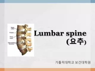
脊柱的 CT 诊断 Diagnosis of Spine. 侯 键 Hoj@sina.com. 第一节 检查方法. 1. SURVIEW 2. SCAN ANGLE 3. SLICE WIDTH 4. WINDOW TECHNIQUE 5. TARGET TECHNIQUE 6. OBLIQUE 7. ANGIOGRAPHY & C OMPUTED T OMOGRAPHIC M YELOGRAPHY -CTM. 扫描定位图象 SURVIEW. SCAN ANGLE 扫描角度. 第一节 检查方法.

E N D
脊柱的CT诊断Diagnosis of Spine 侯 键 Hoj@sina.com
第一节 检查方法 1. SURVIEW 2. SCAN ANGLE 3. SLICE WIDTH 4. WINDOW TECHNIQUE 5. TARGET TECHNIQUE 6. OBLIQUE 7. ANGIOGRAPHY & COMPUTED TOMOGRAPHIC MYELOGRAPHY -CTM
扫描定位图象 SURVIEW SCAN ANGLE 扫描角度
第一节 检查方法 • (2)扫描角度(SCAN ANGLE) • 椎体、椎间盘 • 颈椎、胸椎、腰骶椎 • 扫描方式:standard model
第一节 检查方法 • (3)脊柱CT扫描的层厚(SCAN WIDTH): • 层厚(slice width):椎体、椎间盘、颈椎、腰椎 • 与层距(increment)的区别
(4)窗技术WINDOW TECHNIQUE C1 75 -CENTER W1 250 -WINDOW
第一节 检查方法 • (6)空间重建: • 冠矢状位重建(OBLIQUE) • 与数-摸转换图象重建(RECONSTRUCTION)的区别
第一节 检查方法 • (7)造影检查: • 增强扫描(Contrast) • CTM(Computed Tomographic Myelography)
CTM的部位 硬脊膜 蛛网膜 软脊膜
第二节 正常CT表现 • 脊椎、小关节、椎管 • 椎间盘、椎间孔 • 神经根、神经节 • 脊髓、硬脊膜、硬脊膜囊、蛛网膜下腔、硬脊膜外间隙 • 脊柱的静脉、韧带 • 椎管的测量
脊椎、椎管 颈椎 环椎 C7 C4
胸椎 椎体 椎弓根 骨性椎管 上关节突 椎板 椎板 棘突
腰椎 小关节
椎间盘:髓核与纤维环、透明软骨终板 • 髓核: • 退化的脊索细胞和纤维软骨 • 粘液胶冻样物质 • 纤维环: • 纤维软骨与多层胶原纤维 • 透明软骨终板 • 椎间盘高度: • C-L:3-5mm至15mm
正常椎间盘CT表现: (1)与相邻椎体形状一致 (2)与相邻椎体大小一致 (3)密度均一 (4)软组织密度 CT值:80-120Hu (5)髓核与纤维环不能区分 (6)颈段近圆形 胸段后缘深凹 腰段后缘浅凹 骶段后缘平直或后凸
THE LATERAL RECESS A small, lateral bony excavation can be seen on the vertebral body, especially near the last three lumbar segments. The nerve root passes through the lateral recess before it enters the intervertebral foramen from a point below the root of the arch. The width of the recess :larger than 5 mm It is assumed to be constricted at widths of 3 mm or less.
ligamentum flavum the superior articular process
脊神经节 椎间孔 椎间盘碎片坏死
脊髓 蛛网膜下腔
颈髓 胸髓
马尾与终丝 腰髓 脊髓圆锥
硬脊膜外间隙:脂肪、神经、淋巴、血管、结缔组织硬脊膜外间隙:脂肪、神经、淋巴、血管、结缔组织
硬脊膜外间隙 神经根鞘
脊柱的静脉 椎体静脉
椎体静脉 椎前内静脉 根静脉
椎管内韧带 椎间盘 后纵韧带 黄韧带
椎管的测量 1.Bone window 2.Lumbar slice AP:C:11mm,L:12mm Lumbar canal
编写与制作 成都中医药大学附属医院 摄影:侯键 云南丽江