Download
1 / 69
1.12k likes | 3.33k Views
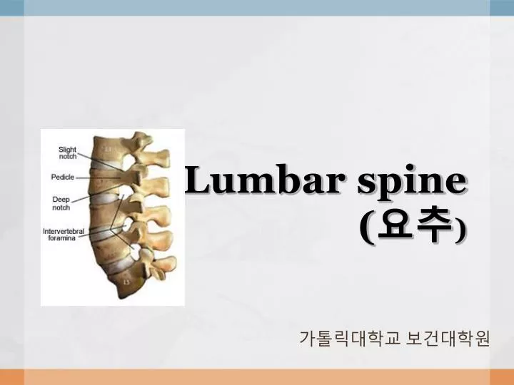
Lumbar spine ( 요추 ). 가톨릭대학교 보건대학원. Applied Anatomy. 운동절의 손상 , 퇴행성 변화 , 외상으로 인하여 … Spondylosis ( 척추협착증 ) degeneration of the IVD Spondylolysis ( 척추분리증 ) vertebra 의 arch 나 pars interaticularis 에서의 defect 모양이 강아지처럼 생겨서 scottie dog 라고도 함 과도한 extension 시에 발생 가능 ( 체조 선수 )

E N D
Lumbar spine(요추) 가톨릭대학교 보건대학원
Applied Anatomy • 운동절의 손상, 퇴행성 변화, 외상으로 인하여… • Spondylosis (척추협착증) • degeneration of the IVD • Spondylolysis (척추분리증) • vertebra의 arch나 pars interaticularis에서의 defect • 모양이 강아지처럼 생겨서 scottie dog라고도 함 • 과도한 extension 시에 발생 가능 (체조 선수) • 나이에 상관없이 다양한 연령대에서 발생 가능 • Spondylolisthesis (척추전방전위증) • 하나의 vertebra가 다른 vertebra 위에서 전방으로 displacement • L5가 S1 위에서 전방으로 나오는 것이 가장 흔함
Applied Anatomy • Lumbar spine • Resting position : midway between flexion & extension • Close packed position : extension • Capsular pattern : side flexion & rotation equally limited, extension • IVD( intervertebral disc ) • annulus fibrosus : fibrocartilage로 구성 & 약 20겹으로 나이테 모양처럼 둘러싸여 있음 • nucleus pulposus : 처음에는 85~90%의 수분을 함유 → 노화함에 따라 65%까지 감소 (탄력성 저하) • Herniations of IVD • Activity & Percentage Increase in Disc Pressure at L3 : coughing or straining (5-35%), Laughing (40-50%), walking (15%), side bending (25%), small jumps (40%), bending forward (150%), lift (73-169%)
Herniations of IVD • Post. longitudinal lig.가 상대적으로 약하기 때문에 대부분 뒤로 탈출증이 나타나고 spinal cord와 주변의 신경들을 압박하게 되어 다양한 증상이 나타남 (IVD는 No nerve, avascular)
Herniations of IVD • Protrusion (돌출) • annulus fibrosus가 찢어지지는 않고 약간 disc가 bulge • 약간의 통증이 허리와 엉덩이 부위에 나타남 • Prolapse(탈출) • 가장 바깥쪽의 annulus fibrosus만 nucleus를 포함하고 있는 상태 • 압박되는 신경의 부위에 따라 통증이 다양하게 나타남 • Extrusion (유출) • disc herniations (annulus fibrosus disrupted) • epidural space로 들어가게 됨 : spinal cord는 epidural space → dura matter → subdural space → arachnoid → subarachnoid space (CSF가 순환) → pia matter 순으로 둘러싸여 있음 • Sequestration (분리) • free nuclear material • cauda equina syndrome 유발→신경압박, 배뇨장애, 지속적인 신경문제 발생
Patient History • 환자의 연령 • 직업 • 성별 • 통증의 원인(손상기전) • 통증 발생 시기( 3-4주 : acute / 12주까지 지속 : subacute / 3년 이상 지속 : chronic) • 통증 부위 • 방사통 (Fig 9-11) • 통증 양상 • 통증의 호전 ,악화 여부 • 기침,웃음,심호흡시 통증 증가 여부 • 통증 악화시키는 자세,동작 & 통증이 완화되는 동작 • 하루 일과 중 통증이 더 심해지는 지 여부 • 이상감각, 무감각증 유무(Fig9-14) • 근력감소나 약화 유무 • 기타- 습관적인 동작 ,수면시 자세, 배뇨 장애, 약물 치료 유무, 일상생활활동과의 관계
Centralization & Peripheralization • Centralization: 통증이 말단에서 점진적으로 중앙에 몰리는 형태 • Peripheralization: centralization 에 대한 반대 개념. 중앙에서 점점 말단으로 통증이 퍼져 나감 더 악화된 상황을 나타내는 것이 대부분임.
Possible effects of disc herniation • L4-L5 사이에서 발생시 5th lumbar root를 압박 • L5-S1 사이에서 발생시 1st sacral N.와 5th lumbar N. root를 압박 • massive central sequestration of the disc at the L4-L5에서는 cauda equina의 모든 신경근을 압박하고 bowel & bladder paralysis 유발 가능 • Herniations는 주로 posterolateral 방향으로 잘 일어남
Observation • Ant./Post./Lat. View(Fig.9-15)
Observation • Markings(피부상태) • Congenital scoliosis & diatematomyelia (선천성 척수 갈림증) • spina bifida occula와 같음 • scoliosis와 동반되는 경우가 많음 • 어깨가 한쪽으로 내려간 것을 관찰 가능 • hairy 가 나타남 • Neurofibromatosis (신경섬유종증) :left thoracolumbar scoliosis가 동반
Step Deformity • spondylolisthesis를 나타냄 • vertebra가 아래의 것보다 전방으로 밀림 → 부러지면서 spinous process가 뒤로 튀어나옴 <Fig. 9-20>
Examination • Active movements (Fig. 9-21) • Flextion : 40-60° • Extension : 20-35° • Side flexion : 15-20° • Rotation : 3-18°
Examination • Sphinx position (Fig 9-24) • 환자의 팔꿈치를 바닥에 대고 손으로 턱을 받쳐 척추를 과신전시킨 후 복부의 힘을 완전히 뺀다. • Quick test (Fig 9-29) • 쪼그려 앉았다 일어났다하는 동작을 빠르게 반복 • 하지의 관절 검사
Herniated disc problems • L4-L5 & L5-S1 사이에서 가장 호발 • nerve irritation, siatica 발생 • lateropost.로 튀어나온 경우 반대방향의 몸을 lat. Shift • centropost.로 튀어나온 경우동측으로 몸을 lat. shift 하는 compensation 이 일어남
Trendelenburg test • gluteus medius 이상이 있을 때 손상측 한쪽 발로 지지시에 반대쪽 pelvic이 내려감 • 이에 대해 compensation으로 trunk가 환측으로 side flex
Resisted Isometric Movements of the Lumbar Spine : 일어나는 움직임은 동일 & 환자에게 저항을 가하는 자세는 thoracic과 동일 • Muscles ① Erector spinae group (최장근, 장늑근, 극근) ② Semispinalis : rotators, multifidus ③ Deep spinalis : ⓜ spasm에 의한 Back pain 발생 가능 특히 extensor ⓜ에서 잘 발생함
Dynamic abdominal endurance test : 복근의 지구력을 검사하도록 고안. : 누운 자세에서 양 손을 옆에 두고 고관절 45˚,슬관절 90 ˚굴곡 -> 턱을 당기고,체간을 구부려 손가락 끝이 선에 닿도록 반복시킨다.
Dynamic extensor endurance test : 엎드린 자세에서 손을 머리 뒤, 양 옆으로 하고 trunk extension
Int./Ext./abdominal oblique test : 양손을 옆으로, 어깨에, 머리 뒤에 놓고 대각선방향으로 윗몸 일으키기
Dynamic horizontal side support : 팔꿈치를 대고 옆으로 누운 자세에서 무릎 or 발/발목을 지지하며 pelvic 들어올림
Back rotators/multifidus (Semispinalis : intermediate에 위치) • 손과 무릎을 대고 엎드린 자세 • 한쪽 팔만 들어 올림 • 한쪽 다리만 들어 올림 • 한쪽 팔과 반대쪽 다리를 동시에 들어 올림
Myotomes L2 : hip flex / L3 : knee ext. / L4 : ankle dorsiflex. / L5 : G. toe ext./S1: ankle plantar flex. ankle eversion, hip ext./ S2: knee flex.
Test for neurological Dysfunction (Neurodynamic Tests) • Slump Test (Fig. 9-50) → thoracic 에서도 적용 • 환자는 table 가장자리에 앉아 • 손을 등 뒤로,고개를 앞으로 당김 (slump lumbar & thoracic spine) • 검사자는 어깨를 아래로 누름.
Slump Test(2) • 환자는 head flex. • 검사자가 cervical에 조심스럽게 overpressure • 검사자가 피검사자의 knee를 조심스럽게 ext.후 발을 dorsiflex시킴. • 환자는 head를 ext. • 양성반응: 무릎을 완전히 편 상태에서 목을 신전시키면 증상이 감소되고 목을 굴곡시키면 다시 증상 증가
Well leg raising test of Fajersztajn : unaffected leg를 들었을 때 affected leg에 통증이 나타나는 것은 다리를 올릴 때 반대쪽 신경이 늘어나면서 병변부의 N. root가 눌려서 나타나는 것임(Fig.9-57)
Prone Knee Bending (Nachlas) Test • 환자를 엎드리게 한 후 수동적으로 가능한 범위까지 knee flex • 검사자는 환자의 hip이 rotation 되지 않았는가 확인함 • unilateral neurological pain → lumbar. buttock. post. thigh→ L2 of L3 N. root lesion • pain → tight quadriceps ⓜ or stretching of femoral N. • Lat. femoral cutaneous N, Saphenous N. 검사 가능 (Fig 9-58, 9-59)
Brudzinski - Kernig Test • supine 손을 머리 뒤로 → flex. head • hip flex & knee flex → raise the extended leg actively (통증이 느껴질 때까지) 양성반응: meningeal irritation, n. root involvement, Duralirritation의미
Naffziger's Test : jugular vein을 약10초동안 누름 → 환자얼굴이 붉어지면 기침하라고 함. 양성반응: 요통 발생 CFS(subarachnoid)의 압력 증가의미
Valsalva Maneuver : 환자에게 앉은 자세에서 숨을 크게 들어마신 상태를 잠시 유지하며 마치 변을 보는 것처럼 힘을 주라고 함. : 통증-> 척수막내 압력 증가 의미
Femoral n. traction test : hip & knee extension → knee flexion traction → neurological pain radiate down ant. thigh
Bowstring test : SLR test 후에 (양성반응일 경우 실시) 같은 자세에서 시작 → popliteal area를 누름 → Sciatic n. 압박 증가 → SLR과 같은 반응이 나타남
Test for Lumbar instability • H & I stability tests • ⓜ spasm & instability detect 검사 • side flex. → side flex & forward flex. → side flex. & ext. → forward flex. → forword flex & side flex → ext. → ext. & side flex • Hypomobile segment가 있으면 최소한 운동의 2곳에서 제한 • Instability가 있으면 one quadrant을 침범하여 운동의 한곳에서만 제한
Specific Lumbar Spine torsion Test • right side lying & lumbar 약간 ext. • rot. & side bending • 검사자는 오른팔을 잡고 L5 spinous proc.에서 upward. Forward 로 pull하면서 움직임 촉진 • L5 위는 고정
Farfan Torsion Test • nonspecific test → facet Jt, Jt capsule, supraspinous/ interspinous lig. neural arch. longitudinal lig. disc • 환자는 prone position & 검사자가 ribs & spine 고정 → 손을 ilium 앞쪽에서 잡고 뒤로 당김 → spinerotation (Opposite side)
Lat. Lumbar Spine Stabiliy Test : 환자를 side lying 후검사자는 L3 정도의 thoracic에 forearm을 놓고 downward pressure (transverse proc. 방향으로)
Test of Ant. Lumbar Spine instability • 환자 side lying & hip flex to 70°& knee flex • 검사자는원하는 극돌기부위에서 촉진 • 피검자의 무릎을 뒤로 밀기 (femur를 따라서) < Fig.9-69 >
Test of Post. Lumbar Spine Instability 환자를 edge에 seat 검사자는 환자의 앞, 허리를 안고 환자가 팔을 구부려 검사자의 어깨에 대고 → 검사자는 환자의 팔을 lumbar 쪽으로 밈. ( lordosis 생성 & sacrum 고정) • L5-S1에 stress & 이를 유지하면서 환자에게 팔로 검사자를 밀도록 지시
Segmental Instability Test • prone & 다리를 내리고 or 다리를 들고 →검사자가 환자의 lumbar spine(post)에 pressure →수동적으로 knee flex. → spine이 hyperextension 되면서 pain ↑
Tests for Joint dysfunction • One - leg standing (Stork standing) Lumbar Ext. Test (Fig 9-73) :환자는 한 다리로 서서 spine을 ext. 하면서 balance 유지 → pain : positive <- Quadrant Test (Fig 9-74) : 환자는 검사자 앞에 서서 spine ext. & side flex. → pain : positive
Mckenzie's Side Glide Test (Fig 9-75) : 환자의 pelvis를 옆에서 감싸 안고 glide → neurological symptoms (affected side)
Tests for intermittent Claudication • Bicycle Test of van Gelderen (Fig 9-76) • sitting erect & sitting flexed 로 자전거 타기 • 엉덩이와 대퇴 후면에서 통증 나타나거나 tingling : positive
Tests for Malingering (꾀병환자) • Hoover test (Fig 9-77) • 정상적으로 한쪽 다리를 올리려고 할 때 → 반대쪽 다리는 아래로 내리려고 함 • 약한 다리를 올리려고 할 때는 → 반대쪽 다리가 아래로 내려가지 않음
Burns Test (Fig 9-78) • 무릎을 꿇고 의자에 올라가서 손을 바닥에 댐 → positive = malingering : unable or overbalance
Reflexes & cutaneous Distribution • Patellar (L3-L4) / Med. hamstring (L5-S1) / Lat. hamstring (S1-S2) / Post. tibial (L4-L5) / Achilles (S1-S2)
Superficial cremasteric reflex (Fig. 9-80) • 날카로운 것으로 안쪽 대퇴를 stroking • negative reflex : scrotum's rising on that side • 반응이 없을 경우 UMN lesion • Superficial abdominal reflex (Fig 9-81) • 날카로운 것으로 복부 stroking • reflex는 skin movement • 반응이 없으면 UMN lesion • unilateral로 없을 경우에는 LMN lesion (T7-L2)