Download
1 / 16
160 likes | 246 Views
Manifestation of Novel Social Challenges of the European Union in the Teaching Material of Medical Biotechnology Master’s P rogrammes at the University of Pécs and at the University of Debrecen Identification number : TÁMOP-4.1.2-08/1/A-2009-0011.

E N D
Manifestation of Novel Social Challenges of the European Unionin the Teaching Material ofMedical Biotechnology Master’s Programmesat theUniversity of Pécs and at the University of Debrecen Identificationnumber: TÁMOP-4.1.2-08/1/A-2009-0011
Manifestation of Novel Social Challenges of the European Unionin the Teaching Material ofMedical Biotechnology Master’s Programmesat theUniversity of Pécs and at the University of Debrecen Identification number: TÁMOP-4.1.2-08/1/A-2009-0011 Dr. PéterBalogh and Dr. Péter Engelmann Transdifferentiation and regenerative medicine – Lecture 6 Hematopoietic stemcells andtransdifferentiation
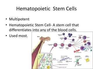
Issues of hemopoietic differentiation • Development of hemopoietic system • Embryonic • Postnatal • Regenerating hemopoiesis • Use of HSCs in non-hemopoietic regenerative medicine
Ontogeny of embryonic hemopoietic tissues Vitelline artery Chorion YS blood islands Allantois Placenta Yolk sac Placenta Umbilical artery Umbilical artery Vitelline artery Liver AGM Allantois Yolk sac Liver Embryo pSP AGM E7.5 E8.25 E9.0 E10.5 Hemangioblast Hemogenic Endothelium Primitive Pro-definitive Meso-definitive Meta-definitive Adult-definitive Myeloid Lymphoid-Myeloid CFU-s Neonatal HSC HSC
Evolution of hemopoietic tissues in rodents Yolk sac Allantois Placenta Para-aortic Splanchnopleura AGM Aortic clusters E7.5 E8.5 E9.5 E10.5 E11 E11.5 E12.5 E13.5 E14.5 E15 birth Primitive Pro Meso Meta Adult Liver Onset of circulation Thymus Spleen Bone marrow
Characteristics of murine embryonic HSCs (AGM/YS/FL) • Leukocyte surface markers: Ly-6A(Sca-1), c-kit+, CD34+, CD45+, • Shared endothelial markers: CD31+, VE-cadherin+ • TF:Runx1+ SCL+ Gata-2+
Transcriptional induction of eHSCs • Intrinsic signals: TF • Runx1: promotes fetal transition of hemogenic endothelium into hemopoietic cells • GATA-2:sequential promotion of mesodermal specification, hemangioblast formation and erythroid differentiation
Extrinsic regulation of eHSCs • Extrinsic signals: interactions with other germ layer elements • Yolk sac (endoderm and mesoderm) • The chorio-allantoic placenta (mesoderm and trophectoderm) • AGM region (dorsal ectoderm, mesoderm and ventral endoderm) • Ventralizing factors– promote hemopoiesis (VEGF, bFGF, TGFβ and BMP4) • Dosalizingfactors– antagonize hemopoiesis (EGF and TGFα)
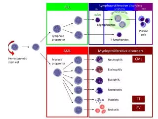
Hemopoietic differentiation models M Classic dichotomy model Myeloid-based model T M T B M T B B (CMEP) (CMEP) M T M E m T B E m M E m T B E m T B M E m T B M E m E m M E m T B M E m (CLP) M B M M M Modified classic model M M M M (CMLP) (CMEP) T T Myeloid potential M Erythroid potential B B E Megakaryocyte potential m T-cell potential (CMLP) T (CLP) B-cell potential B
Transcriptional regulation of early hemopoietic commitment Hemangioblasts Hemogenic endothelium SCL AML-1 GATA-2 Lmo HSC Notch1 Ikaros HoxB4 GATA-2 p21 PU.1/GATA-1 PU.1/GATA-3/Ikaros Bcl-2 CMP CLP Apoptosis
Transcriptional regulation of myeloid differentiation Monocyte ICSBP, PU.1 Neutrophil HSC PU.1 C/EBP C/EBP, GATA-1 PU.1 & GATA-1 Eosinophil GATA-1 GATA-1/FOG Erythrocyte EMP CMP GMP GATA-1, 2 Megakaryocyte
Transcriptional regulation of lymphoid differentiation HSC Notch1 PU.1 IL-7R Pre- pro-B D-JH Monocyte V-D-JH PU.1 GM-CSFR Pu.1, E2A Pax5? EBF Pax5 Pro-T CLP Early pro-B Late pro-B B cell
Steady-state and activatedhaemopoiesis Endothel Fibroblast SCF, FLT-3I, TPO EPO G-CSF, GM-CSF Osteoblast HSC HSC HSC HSC HSC HSC HSC HSC Anemia Hypoxia Blood vessel Endothel EPO Fibroblast G-CSF, GM-CSF TPO IL-1 TNF TGF Osteoblast Macrophage Bacterial infection Inflammation
Human hemopoietic potential • Intraembryonic sources and potential: • D19: HPP in embryo in theabsence of detectable CD34+hematopoietic cells, andspanned both lymphoid and myeloid lineages • D24: in the splanchnopleural mesoderm • D27:aorta with CD34+ progenitors • Yolk sac: only myelopoiesis starting at around the 3rd week
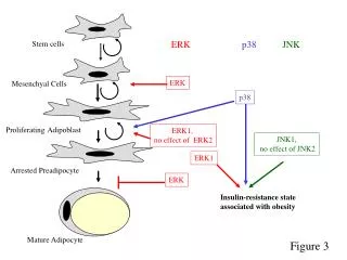
Other potential uses of hemopoietic stem cells • Regenerative medicine in parenchymal tissues: muscle, neural tissues, liver, etc. • Sources: adult or embryonic (umbilical vein mononuclear cells) • Experimental settings: use of genetically marked cells or inducible Cre-Lox transgenic animals, and their detection in damaged/regenerating tissues
Summary • Hemopoiesis is established in successive waves at various anatomical locations, where hemopoietic activities at different host tissues result in diverse cellular progeny. • The hemopoieticcommittment is under the combined effects of endogenous programing and external signals, where latter elements may alter the steady-state hemopoiesis. • Hemopoietic stem cells may promote the regeneration of non-hemopoietic tissues by (a) promoting vascular repair, (b) tissue repair and (c) possible transdifferentiation.