Download
1 / 85
940 likes | 1.27k Views
Chest Trauma. General Aspects of Chest Trauma. 25 % of trauma deaths are due to chest injury 85 % are treatable by simple procedures Only 15 % or less require thoracotomy for repair Mechanisms of injury: Penetrating Blunt Blast Inhalation (fumes, water, etc.).

E N D
General Aspects of Chest Trauma • 25 % of trauma deaths are due to chest injury • 85 % are treatable by simple procedures • Only 15 % or less require thoracotomy for repair • Mechanisms of injury: • Penetrating • Blunt • Blast • Inhalation (fumes, water, etc.)
Potential Physiologic Results of Chest Trauma • Hypoxia • Hypercarbia • Hypovolemic shock • "Obstructive" shock • Acidosis
Information from prehospital personnel, such as description of this type of steering wheel damage, can heighten suspicion for significant chest injury
Chest Trauma • The 6 quickly lethal types of chest injury : (these must be found on the primary exam) • Airway obstruction • Tension pneumothorax • Open pneumothorax • Massive hemothorax • Flail chest • Cardiac tamponade
Chest Trauma • The 6 potentially lethal types of chest injuries : (these should be identified on the secondary survey) • Aortic disruption (dissection) • Myocardial contusion • Tracheobronchial disruption • Esophageal disruption (perforation) • Pulmonary contusion • Diaphragmatic disruption (hernia)
The 8 Types of (Usually) Non-Lethal Chest Injuries • These should be identified on the secondary survey : • Simple pneumothorax or small hemothorax • Sternoclavicular dislocation • Sternal fracture • Clavicle fracture • Scapular fracture • Traumatic asphyxia • Simple rib fracture • Chest wall contusion
Quickly Lethal Chest Trauma : Airway Obstruction • Should diagnose this "from across the room" by observing : • Decreased respiratory effort or rate < 12 / min. • Cyanosis • Intercostal / sternal / subcostal retractions • Snoring / gurgling / hoarseness / stridor • Agitation or obtundation
Quickly Lethal Chest Trauma : Airway Obstruction • Treatment (more detailed review is in the E.T.C. course section on airway) • Oxygen (high flow) • Airway opening maneuvers • Suction • Oropharyngeal or nasopharyngeal airway • Invasive airway management • Endotracheal intubation • Needle or surgical cricothyroidotomy • These should be done as part of the primary survey
Quickly Lethal Chest Trauma : Tension Pneumothorax • Signs : • Respiratory distress • Trachea deviated to opposite side • Breath sounds decreased or absent on injured side • Expansion or hyperinflation of injured side • Tympanitic percussion on injured side • Often neck veins distended
Quickly Lethal Chest Trauma : Tension Pneumothorax • Treatment : • Do not wait for CXR confirmation • Immediate decompression with 18 to 14 gauge needle inserted in 2nd intercostal space in mid-clavicular line along upper edge of rib : allows air under pressure to escape to relieve the tension • Should then insert thoracostomy tube to water seal • Should do these as part of primary survey
Quickly Lethal Chest Trauma : Open Pneumothorax • Occurs if open hole in chest wall is > 2/3 the diameter of the trachea (air flow via trachea is reduced) • Treat by placing an occlusive dressing of vaseline gauze taped down to skin on 3 sides (if taped on 4 sides can lead to tension pneumothorax) : allows vented air to escape • Definitive treatment is surgical debridement and closure of the chest wall defect and concurrent tube thoracostomy
Quickly Lethal Chest Trauma : Massive Hemothorax • Represents > 1500 cc blood in pleural cavity • Signs : • Shock • Flat neck veins • Decreased breath sounds on injured side • Dullness to percussion on injured side
Quickly Lethal Chest Trauma : Massive Hemothorax • Treatment : • Timing of chest tube placement is a tricky decision : if too early, bleeding point may untamponade and patient can exsanguinate • Usually best to start to restore intravascular volume before chest tube placed • Send type & cross and have blood ready • Be ready to do thoracotomy • "Cell saver" for autotransfusion may be very helpful
Quickly Lethal Chest Trauma : Flail Chest • Occurs if > 3 ribs are fractured in 2 or more places • Results in a "free-floating" section of chest wall and paradoxical respiratory phase movement of the flail segment, contributing to ventilatory insufficiency • Underlying pulmonary contusion is often present • Very seldom requires any type of surgical therapy
Quickly Lethal Chest Trauma : Flail Chest • Often can treat this "conservatively" with broad taping of the flail segment, oxygen, fluid restriction, suction, intercostal blocks • Consider early treatment with intubation, mechanical ventilation + PEEP if : • Age > 65 yrs • Other major injuries present • PCO2 elevated (> 44 mm Hg) • PO2 decreased (< 60 on 40 % O2 by face mask) • Subjective resp. distress or increased resp. rate • Pre-existent COPD
Quickly Lethal Chest Trauma : Cardiac Tamponade • Diagnose by Beck's Triad : • Hypotension • Distended neck veins (JVD) • Muffled heart tones • Note : Tension pneumothorax & cardiac tamponade are the only acute traumatic causes of shock with distended neck veins
Quickly Lethal Chest Trauma : Cardiac Tamponade • Treatment : • Nonsurgical measures can be used to temporize most patients till a chest surgeon is available • IV fluids : push CVP to 18 to 20 cm. H2O • Sometimes dopamine drip (2 to 10 micrograms / Kg / min.) helpful • Consider needle pericardiocentesis (risk of coronary artery or ventricular wall injury) • Consider subxiphoid pericardial window under local anesthesia • Thoracotomy and repair of cardiac injury is definitive Rx
Potentially Lethal Chest Injuries : Aortic Disruption (Rupture) • Major cause of death from MVA's or falls from a height • CXR signs : • Wide mediastinum (> 8 cm on AP view at level of aortic knob) • Blurring or obliteration of aortic knob • Left pleural cap +/- left pleural effusion • Deviation of trachea or NG tube to right • Depression of left mainstem bronchus • Separation of calcified aortic plaque from aortic edge > 5 to 6 mm • Other signs : • Pulse deficit or BP difference between arms • Paraplegia • Lower extremity hypotension
Potentially Lethal Chest Injuries : Aortic Disruption • Sites of rupture : • 80 to 90 % just distal to takeoff of left subclavian (at ligamentum arteriosum) • Remainder are at aortic root or diaphragm • Confirm diagnosis : • Angiography is "gold standard" but spiral computed tomography (CT) with contrast has been shown very sensitive and accurate • Recent reports indicate transesophageal echocardiography (TEE) is highly accurate (but is operator dependent) • Non-spiral CT of chest may miss up to 30 %
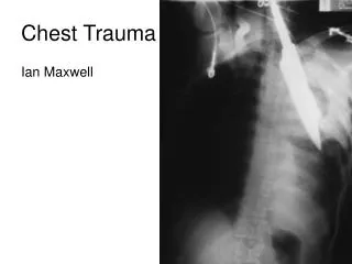
Posterior dislocation of the manubrium causing wide appearance of the mediastinal shadow on chest X-ray
Potentially Lethal Chest Injuries : Aortic Disruption • Treatment : • Avoid "over-resuscitation" and hypertension (more likely to uncontrollably rupture if BP > 140 / 90) • Type and cross for at least 10 units of blood • To OR emergently for surgical repair (usually synthetic graft interposition required) • Only pre-emptive surgery is laparotomy for active bleeding in abdomen ; then do thoracotomy & aortic repair after laparotomy
Potentially Lethal Chest Injuries : Pulmonary Contusion • Signs : • Hemoptysis • Decreased breath sounds • Dullness on percussion • Respiratory distress • Hypoxemia • Infiltrate on CXR • Often associated with rib fractures
Potentially Lethal Chest Injuries : Pulmonary Contusion • Treatment • Oxygen • Pulmonary toilet • Restrict fluids • Bronchodilators only if wheezing • Steroids contraindicated • Antibiotics not helpful initially • Follow with daily serial CXR's, +/- ABG's, +/- PFT's
Chest X-ray showing fracture of left clavicle, right pulmonary contusion, and right pneumothorax
Potentially Lethal Chest Injuries : Tracheobronchial Disruption • Due to major laceration in trachea or bronchus • Diagnose by large air leak in chest tube • Often even a second chest tube cannot overcome the air leak & allow reexpansion of the lung • Often have very large amount of subcutaneous air • Treatment • 2nd chest tube to suction • +/- selective endobronchial intubation (Carlens tube) • To O.R. emergently for bronchoscopy + thoracotomy + surgical repair
Potentially Lethal Chest Injuries : Esophageal Disruption (Rupture) • Most common with penetrating injury but can occur from blunt trauma • Signs • Dysphagia • Deep chest pain • Subcutaneous +/- mediastinal air • Pneumothorax +/- pleural effusion • Cloudy fluid or high amylase in fluid from chest tube
Potentially Lethal Chest Injuries : Esophageal Disruption (Rupture) • If suspected : • Gastrografin swallow or esophagoscopy • Place chest tube as soon as possible to control drainage • Broad spectrum antibiotics • If confirmed : • To OR emergently for repair or esophageal diversion
Potentially Lethal Chest Injuries : Diaphragmatic Disruption (Rupture) • Risk is herniation of abdominal viscera into chest with strangulation • Also can result in lung compression & ventilatory compromise from viscera in chest • If initially missed can present with complications even years later
Potentially Lethal Chest Injuries : Diaphragmatic Disruption • Suspect the diagnosis if : • CXR shows elevation or indistinctness of one hemidiaphragm • CXR may show dense basilar infiltrate • Diagnosis confirmed by : • CXR showing bowel, stomach, or NG tube in chest • Peritoneal lavage fluid exiting from chest tube • Computed tomography of lower chest • Sometimes need gastrografin UGI • Treatment : • To O.R. for laporatomy for repair • NG tube preop to decompress stomach