Download

1 / 59
600 likes | 645 Views
RECURRENT MISCARRIAGE CURRENT CONCEPTS. SUSHANTA BHADRA FEBRUARY 2004 WEXHAM PARK. DEFINITION. Loss of three or more clinically recognized pregnancy losses before 20 wks gestation.

E N D
RECURRENT MISCARRIAGECURRENT CONCEPTS SUSHANTA BHADRA FEBRUARY 2004 WEXHAM PARK
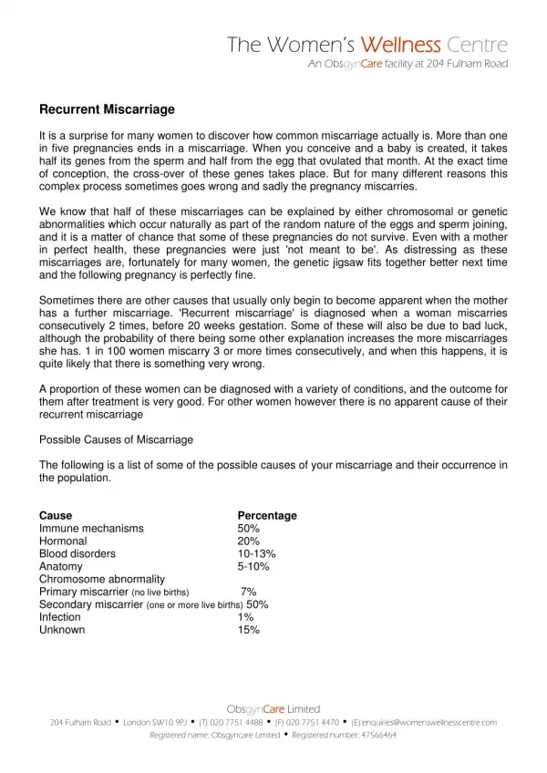
DEFINITION • Loss of three or more clinically recognized pregnancy losses before 20 wks gestation. • Clinical investigation should however be initiated after 2 consecutive losses specially when fetal heart activity has been identified before any pregnancy losses, when the woman is >35 yr or when the couple has difficulty conceiving.
epidemiology • Affects 0.5-3% of all women • Risk of subsequent loss - 24% after 2 30% after 3 40% after 4
PROPOSED ETIOLOGIES • Genetic - 5 % • Anatomic- 15% • Endocrine- 20% • Infections- 5% • Immunologic/ Thrombotic -30% • Other factors – 10% • Unknown
Genetic mechanisms • Chromosomal abnormalities numerical – aneuploidies structural - translocations • Single gene ( Mendelian) • Polygenic ( single anatomic defect)
Chromosomal abnormalities Spontaneous abortions Normal chromosomes – 40-50% Abnormal chromosomes- 50-60%
aneuploidies • Trisomies (extra chromosome) and monosomies (missing chromosome) • Segregation errors during cell division • Sporadic • Nonrecurrent • Trisomies associated with maternal age
Abnormal chromosomes • Autosomal trisomy –50% • Monosomy X – 25% • Polyploidy –20% • Sex chromosome polysomy - rare • Translocations - < 5%
Autosomal trisomies • Chromosomes 1—0% 13– 5.8% 2—5% 14, 18 —5% 3,5,6 ,11,12,17 - <1% 15—7.2% 4 , 20 –2.5% 16—31% 7—4.5% 21 – 8.4% 8,9—3.5% 22 ---11%
Parental origin - trisomy • Maternal –90—95% -- age related recurrent • Paternal – 5—10%
Aneuploid screening • There is an increased rate of numerical chromosomal abnormalities in human periimplantation embryos in women with RSA • There is also an increased incidence of chromosomal abnormalities in the sperm from RSA couples • Role of preimplantation genetic diagnosis (day3 - blastomeres ) using FISH
Structural chromosomal abnormalities • Defect in structure of 1 or more chromosomes • Inversions, translocations • 7% couples affected • Risk of spontaneous abortions vary from 25-50% • May be passed from parent to child • Karyotype indicated
TRANSLOCATIONS • Reciprocal --- any chromosome • Robertsonian –(centric fusion) only acrocentric chromosomes 13,14,15,21,22 • Cryptic translocations - balanced translocations involving only the telomeric regions of the chromosomes – not detectable by conventional cytogenetics
Other chromosomal rearrangements • Inversions • Balanced complex translocations • Interchromosomal insertions • Jumping chromosomes
X Chromosome inactivation • Occurs in female mammals • Random inactivation of a X chromosome to compensate for the difference in x linked gene dosage • Preferential inactivation of x chromosome is directly correlated with RM • Underlying causes include cryptic x chromosome aberrations, gene microdeletions, gene mutations and genetic imprinting
Chromosomal causesConclusions • Aneuploidies are responsible in 55-85% of EPL • Trisomies are usually maternal meiotic in origin and age related. • Polyploidy(67%) and Monosomy X(80%) are usually paternal in origin • Trisomies can be recurrent • Parental translocations found more often in female, not highly correlated with number of losses and show 2-5% unbalanced offspring
Single gene defects • Maternal – endometrial, immunologic, vascular • Embryonic – developmental • Genes conferring pharmacologic susceptibility to toxins or infections • Genes causing aneuploidy
Polygenic • 2 or more genes cumulatively affect presence or absence of a given trait • Unequivocal relationship to 2nd and 3rd trimester losses • Associated with anatomic defects involving single organ system • Associated with subsequent live born ntd and prior polygenic defects • Fetuses with anatomic defects (embryoscopy) usually show cytogenetic abnormalities • Recurrence risk 1-5% limited to first degree relatives
Maternal gene perturbations Mutant maternal gene likely to be associated with consecutive losses not interspersed as in genes acting through embryos • Endometrial receptivity (PR) • Luteal Function (CYP 17) • Alloimmune (HLA G promoter polymorphism)
Lethal genes affecting fetus • Early lethal – Surf 1 , ETA2 , OCT 4( mice models – human analogies present – neurodevelopmental problems ) • Placental – trophoblast differentiation , fetoplacental vascular development , trophoblast transcription factors • Homebox and other developmental : HOX PAX
Hla genotypesThe REMIS Trial • analysis of 12 HLA g alleles in prospectively followed cohorts of couples with recurrent miscarriages using PCR sequence specific oligonucleotides for 12 alleles • 113 couples studied- 63 with successful pregnancy, 50 with rm
Remis trial • HLA g gene genotype 0104 and 0105n is predictive of low successful pregnancy rates • Presence of HLA G isoform 1 and 725C/G polymorphism in promoter regions are associated with an increased risk of recurrent miscarriages if both partners carried the allele
The Paternal contribution • Balanced structural chromosomal abnormalities • Sperm abnormalities • Sub chromosomal abnormalities • subtle chromosome rearrangements • gene dosage imbalances • Mutations
Sperm abnormalities • 24 couples with rm – semen analysis&Fish Characteristic rec misc fertile donors Motile 46% 49% Tapered 38% 16% Amorphous 9% 5% Viable 56% 71% (Carrell 2003)
Sperm abnormalities Disomy rec misc sperm donors Xy 0.77% 0.31% 13 1.02% 0.39% 18 0.51% 0.25% 21 0.47% 0.28%
Sperm aneuploidy • Mechanisms • Quality marker ? • Carrier of a defect that influences post zygotic aneuploidy , implantation, embryonic growth
The role of the trophoblast • Placental development • Continuous turnover • CT proliferation – differentiation – fusion – aging – shedding as syncitial knots into maternal circulation over 3- 4 weeks • CT / ST ratio – reduced in apl pregnancies and rsas • Tenney Parker changes
Placental oxidative stress • Human fetus develops in a low oxygen environment • Intraplacental oxygen conc increases from < 20mm Hg at 10 wks to > 50 at 12 wks • Trophoblastic cells are extremely sensitive to oxidative stress • Mounting evidence that in most miscarriages the onset of intervillous circulation is premature and widespread due to incomplete transformation of uteroplacental arteries leading to high oxygen concentrations in early pregnancy
Endometrial receptivity • INFERTILITY RM • 50-75% of pregnancies lost represent a failure of implantation • Failure of implantation may result from a non receptive endometrium • Involves a complex synchronous interaction between embryo , endometrium and ovary
Endometrial receptivity • Growth factors –LIF,HB EGF • Cytokines • Adhesion molecules- integrins A5, B3 • Steriod hormones and receptors • Immunologic factors-- NK Cells, T cells • Prostaglandins
RX to improve endometrial receptivity • Progesterone—at best controversial , at worst ineffective • Immunomodulation paternal cell immunization intravenous immunoglobulin
Novel Therapies • Intrauterine Prostaglandins • Intrauterine steroids • Intrauterine Peripheral blood mononuclear cells • L arginine • Glue– Fibrin!
Infections • 1 in 20 women are exposed to pathogens • Majority are harmless • Early infection – congenital problems • Delayed infection -
Infections - spectrum • MISCARRIAGES • CONGENITAL INFECTIONS • STILL BIRTH • NEONATAL DEATHS • ASYMPTOMATIC INFECTIONS • NORMAL FINDINGS
INFECTIONS • Rubella • CMV • HBV • VZ • HSV • HIV • GBS • Syph
Infections – What do they do ? • Direct effect on ova • Endometrial infection – implantation defects • Embryopathy • Placental infections • Amniotic fluid infection
UTERINE PATHOLOGY • Septate uterus- • Asherman’s Syndrome- • Uterine Fibroids- esp. sub mucous • Primary endometrial defects • Des exposure
Cervical Cerclage • Shirodhkar • McDonalds • Lash • Benson Durfee
Indication for abd cerclage • Congenital short cx • Amputated cx • Torn cx • Severe scarring • Chronic cervicitis • Cervicovaginal fistula • Failed shirodhkar • Rec pproms • Cervical dysfunction
Cervical cerclage Steer Modifications • Nuchal first • USS guidance – before , during and after • No bladder dissection • Straight blunt needle
PROTHROMBOTIC STATES • Antiphospholipid syndromes • Heritable Thrombophilia-antithrombin def protein C & S def Factor V Leiden Prothrombin20210 A • Thrombocythemia
ANTIPHOSPHOLIPID SYNDROME • 7-42 % OF WOMEN • Wide variation • Poor laboratory standardization
APL – diagnostic criteria • 3 or more unexplained consecutive spontaneous abortions before 10 wks with exclusion of maternal anatomic or hormonal abnormalities and maternal and paternal chromosomal abnormalities OR • One or more unexplained deaths of a morphologically normal fetus at or beyond 10 wks with normal fetal morphology documented by USS or direct examination of the fetus OR • One or more PTBs of a morphologically normal neonate at or before 34wks gest because of severe PET or Placental Insufficiency
AND Persistent abnormality of the following tests when measured twice at least 6 wks apart Lupus anticoagulant Antiphospholipid antibodies – IgG or IgM
Pathophysiology of APS • Thrombotic • Lack of Trophoblastic invasion in 1st trimester decidua
APL – Maternal Risks • Thrombosis –Heparin RX Access to prenatal care and pt education • Hypertension – antenatal care and pt education • Thrombocytopenia • Secondary conditions rheumatologist involvement • Treatment Complications– hge, osteopenia, thrombocytopenia • Catastrophic APS
FETAL RISKS • Miscarriage • Uteroplacental insufficiency IUD IUGR Fetal Distress • Preterm birth • SLE and Thrombosis
Heritable thrombophilias • 5 recognized defects – antithrombin def, protein c def, protein s def, v leiden, prothrombin 20210A variant • EPCOT – European study analysed pregnancy outcome in women with known thrombophilia – v leiden not associated with rm, better association with activated protein c resistance • Essential thrombocythemia
ENDOCRINOLOGICAL FACTORS • Hypersecretion of LH(>10IU/L)In the follicular phase is a marker for RM • Androgen levels in the follicular phase have been shown to be high in pts with RM- This correlates negatively with the conc. of Placental Protein 14 a biochemical marker for endometrial function • Hyperprolactinemia – no firm evidence