Download
1 / 39
390 likes | 393 Views
Water. H-Bonding Angle (linear = strongest) Distance (between donor and acceptor) Partial Charges on Participants Dielectric Constant ( E ) (a measure of a solvent’s ability to shield charges) F = kq 1 q 2 E r 2 Polar: E >15 Apolar: E <15. High Boiling Point

E N D
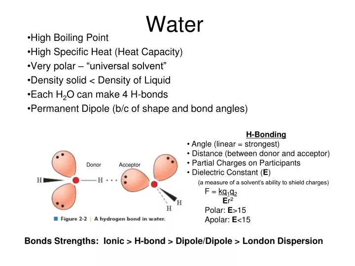
Water • H-Bonding • Angle (linear = strongest) • Distance (between donor and acceptor) • Partial Charges on Participants • Dielectric Constant (E) • (a measure of a solvent’s ability to shield charges) • F = kq1q2 • Er2 • Polar: E>15 • Apolar: E<15 High Boiling Point High Specific Heat (Heat Capacity) Very polar – “universal solvent” Density solid < Density of Liquid Each H2O can make 4 H-bonds Permanent Dipole (b/c of shape and bond angles) Donor Acceptor Bonds Strengths: Ionic > H-bond > Dipole/Dipole > London Dispersion
Acid/Base Chemistry Weak Acid Dissociation: Dissociation Constant Ka = HA H+ + A- HB H+ + B- larger Ka = stronger weak acid smaller pKa = stronger weak acid Henderson-Hasselbach Equation: pH < pKa [A-] < [HA] pH = pKa [A-] = [HA] pH > pKa [A-] > [HA] Can rearrange this eqn to solve for the ratio of deprotonated to protonated: pH – pKa = log[A-]/[HA] pH – pKa = 10^[A-]/[HA] Buffering (80%) occurs +/- 1 pH unit from pKa Best Buffering is at pKa, because here [A-] = [HA] Buffering capacity is the ability of a buffer to resist changes in pH, is dependent on the concentration of buffer and pH of solution
pH = pKa at half equivalence point Can also be OH- equivalents (need 1 mole of OH- equivalents per ionizable group)
Amino Acids • Usually found as a zwitterion • L-stereochemistry Amino group on left Carbon 1 (carboxy) on top
Amino Acid pKa’s • Carboxyl groups pKa ~ 2.0 • NH3 (N-termini) pKa ~ 9.5 • Inductive Effect • e- withdrawing effect • Lowers pKa • Electrostatic Effect • Charge effect • Molecules prefer a net neutral charge • Can raise and lower pKa’s pKa is 2.35 Lower than pKa in acetic acid (CH3-COOH) because N is withdrawing electrons Glycine pKa is 9.78. Higher than NH3 in ethylamine (CH3-CH2-NH3+) because COO- is withdrawing electrons Higher than NH3 in O-methyl glycine (NH3-CH2-C-OCH3) to increase range that glycine has O net 0 charge
Amino Acids Can form an Ion Pair at pH 7: Asp, Glu, Arg, Lys, His (sometimes) Can Disulfide Bond at pH 7: Can participate in Van der Waals contacts at pH 7: ALL 20!
Amino Acids Can H-bond at pH 7: Asp, Glu, Arg, Lys, Ser, Thr, Asn, Gln, Tyr, His, Trp Can Ionize (gain or lose a proton): Charged (Arg, Lys, Asp, Glu, His) Alcohols (Ser, Thr, Tyr) Cys *only side chains with groups that can gain or lose protons can ionize. Note that amino acids with NH2 groups (Asn, Gln) are NOT IONIZABLE!
Isoelectric Point (pI) • Net charge on protein/aa is 0 To solve these problems, make a table with pH ranges that are the pKas. Then figure out the charge on each ionizable group at the given pH. One of these pH ranges will sum to 0. These are the two pKas to plug into the pI equation. pI = (10+12.5)/2 = 11.25
Henderson-Hasselbach Eqn can be used to determine net charge 10pH-pKa = [A-]/[HA] Example: A protein has three ionizable groups (NH3 at N-termini, Arg, Tyr) To find the net charge at pH 7, [NH2]/[NH3+] = 10(7-8) = 10-1= 1/10 10 out of 11 have a +1 charge = 90% [O-]/[OH] = 10(7-10) = 10-3 = 1/1000 1 out of 1001 have a -1 charge = 0.099% [NH2]/[NH3+] = 10(7-12.5) = 10-5.5 = 100% in NH3+ form Add the percentages of each species (paying attention to the sign/charge) +0.9 -0.0009 +1 = +1.9 We can then say that that most molecules have a charge of +2, a few are +1
Condensation Reaction eliminates water, forming a peptide bond that joins two amino acids • Peptide Bond: • Has partial double bond character • Is planar • (N to C) • no at N-terminal • (C to Carbonyl C) • no at C-terminal C-terminus N-terminus
Conformations of the Protein Backbone (, ) are limited by STERIC CLASHES Ramachandran Plot: plots the allowed phi/psi conformations
Determine # of peptide chains present • Count # N-termini • DNFB or dansyl chloride react w/ N-terminus • hydrolyze all peptide bonds (acid treatment) • isolate and ID N-terminal aminos • Problem: reaction at Lys or other 1o amines • 2. Separate Chains • may need to reduce disulfides/ block with IAA • Fragment polypeptides • Enzymatically (endopeptidase) or chemically (CNBr) – these specifically cleave • Sequence Fragments • Edman degradation • Edman’s reagent adds to N-terminal under basic conditions, switch to acidic conditions and cut off N-terminal residue, ID this residue, repeat • Mass spec • Reconstruct sequence • this required fragmenting in different places to get overlapping segments
Multiple sequence alignments • Sequence Identity = fraction of positions that are the same amino acids • Sequence similarity = fraction of positions with the same or similar amino acids (conservative substitutions) • Homologs • Orthologs = proteins of same function but in different organisms • Paralogs = related sequences of slightly different function (same organism) • thought to arise by gene duplication • Conserved and similar positions are probably important for structure/function • Rate of protein mutation is related to the ability of the protein to accommodate the mutation
Rise = 5.4Å (per repeat) 3.6 amino acids per repeat 1.5Å rise per amino acid H-bonding in backbone stabilizes structure C=O of i H-bonds to i+4 Small electric dipole N-termini has free amide groups (+) C-termini has free carbonyls (-) Alpha helices - • Amphipathic helix = half hydrophobic, half hydrophilic • Helical wheel projections can show this • 5 factors influencing helix stability • Intrinsic propensity of amino acids (Ala likes to be in helices) • 2. Interactions between R-groups (ionic interactions) • Bulkiness of adjacent R groups (Phe, Trp) • Occurrence of Pro/Gly (destabilize helices) • Pro is not very flexible and causes helix kinks, • Pro cannot H-bond because its N is missing an H • Gly is very flexible) • Interactions with ends of helix and R groups • (Arg at C-terminal ends) +
Beta-sheet/strand • Antiparallel • 7Å rise • 2 amino acids per repeat • H-bonds are linear • Parallel • 6.5Å rise • 2 amino acids per repeat • H-bonds are slanted • Do not see fully extended (phi = 180o, psi = 180o) because then R groups will start to interfere with protein backbone • Sheets are in non-continuous regions • Beta-turns • 4 amino acids, Pro/Gly common • H-bond b/t C=0 of amino acid 1 and NH of amino acid 4
Stabilizing Interactions in Proteins 1o covalent peptide bond 2o: H-bonding (backbone N-H. . . . O) Electrostatic Ion Pairs Steric compatibility Van der Waals Hydrophobic Effect 3o: 1+2 and disulfide bonds 4o: same as 3o • Hydrophobic Effect • Maximizing the entropy of water • Water is ordered around nonpolar substances. It forms a shell, motion is restricted and entropy is lower • Proteins have a hydrophobic core and a more hydrophilic surface. • This drives protein folding because the protein becomes more ordered but the water becomes less ordered
Carbohydrates (CH2O)n Fisher Projections • If the OH on the last chiral carbon is on the right, sugar is D • If OH on the last chiral carbon is on the left, sugar is L Steroisomers • Number of conf’s possible = 2n (n=# chiral centers) • Epimer = sugars that differ at 1 stereocenter • Glucose and Galactose are epimers at C4 Sugars cyclize • Anomeric carbon • Has 2 bonds to oxygen • Alpha anomer = OH on opposite side of ring as C6 • Beta anomer = OH on same side of ring as C6
Reducing Sugars: • Can reduce Cu++ to Cu+, sugar gets oxidized • Requires the sugar to be linear so that carbonyl is accessible (but remember that cyclic sugars can open up and then be reducing) These are reducing because an OH is attached to the anomeric carbon If the OH was “OR” (a glycosidic bond) then the sugar could not open up and would not be reducing Sugars can mutarotate (interconversion of /β anomers) as long as the sugar is reducing
-Linked Sugars • Loose (flexible), highly hydrated, helical, granular, branched, rings in chair conf • Glycogen • Glucose in 1-4 (linear) and 1-6 (branched) linkages • One reducing end, many non-reducing ends • Chain grows by adding to non-reducing ends • Starch • Amylose (1-4 glucoses, linear) winds in among a mesh of amylopectin • Amylopectin (branched) • Many non-reducing ends, few reducing ends
β-Linked Sugars • Extended, Fibrous, Extensive H-bonding, Rigid, Rings in Chair conformation, Provide Support & Lubrication • Cellulose • Glucose with β1-4 linkages • Extended chains, very close packing, not very much hydration = rigid fibers that are hard to digest • Chitin • β1-4 linked N-acetylglucosamine • Peptidoglycan • Chains of alternating N-acetylglucosamine and N-acetyl muramic acid • Combined with peptides • Rigid mesh-like shell around bacteria • Glycosaminoglycans • Alternating sugar with amino-sugar, β1-3 linkages • Negatively charged • Shock absorbers, highly hydrated • Ex: Heparin
Glycoproteins • N-linked = attached to Asn • Attached during synthesis • O-linked = attached to Ser/Thr • Attached after folding • Microheterogeneity = diversity in sequence of attached sugar • Glycoforms = different patterns of glycosylation
Lipids • Functions: • Energy storage (triacylglycerols) • Membranes (structural) • Signalling • Intracellular (sphingolipids, phosphotidylinositol) • Intercellular • Intertissue (steroid hormones) • Interorganism (pheromones, volatile plant lipids) • Insulation • Light Absorption • Nutrition • Electron Carriers (CoQ) • Enzyme cofactors • Antioxidants
Fatty Acids • COOH at one end, 4-36 carbons • (even # of carbons only) • Lipid oxidation releases energy • Lipids are VERY reduced so they can be oxidized more than sugars • Not hydrated. Means more energy per unit weight • 6x the amount of energy as sugars • Melting Points • Higher as chains get longer (more Van der Waals contacts) • Lower as # of double bonds increases (more kinks = worse packing)
18:2(Δ9,12) or (-6) or (n-6) # Carbons # double bonds Delta name: name starting carbon of the double bond from COOH end • name: name starting carbon of the double bond from methyl end • Double bonds are cis! • Double bonds occur every 3 carbons • We cannot synthesize -6 or -3 FA, need these from diet 18:0 18:1(Δ9) 18:2(Δ9,12) 18:3(-3)
Triacylglycerols • Energy storage, thermal insulation • Naming • For ex: 1-palmitoyl-2-stearoyl-3-oyl glycerol Glycerol-based Lipids: Glycerophospholipids = glycerol + FA + phosphate + group attached to phosphate Glyceroglycolipids = glycerol + FA+ oligosaccharide
Sphingolipids • Sphingosine + FA = ceramide • Sphigomyelins = ceramides with phosphocholine or phosphoethanolamine • Sphingophospholipids (charged)
Glycosphingolipids • Cerebrosides • (monosaccharide attached to ceramide, uncharged) • Gangliosides • (oligosaccharide attached to ceramide, charged, at least one sialic acid attached to sugars
Sterols • Slightly amphipathic because of –OH • Fused planar rings