Download
1 / 69
690 likes | 709 Views
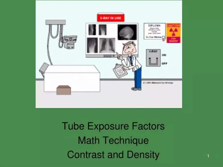
Tube Exposure Factors Math Technique Contrast and Density. X-ray Properties. Artistic Talent. Artistic Talent. Your artistic talent…. Your artistic talent….. Your signature. What is Technique?. Exposure factors under the control of the radiographer at the control panel mAs (mA x s)

E N D
Tube Exposure Factors Math Technique Contrast and Density
What is Technique? • Exposure factors under the control of the radiographer at the control panel • mAs (mA x s) • Milliamperage • seconds • kVp
How does it affect our image?(SLIDE 14) It will tell us if the image is: too dark too light diagnostic
The Control Console (SLIDE 15) • Where tech sets technical factors A. MAS B. KVP C. Makes exposure • Only a legally licensed individual is authorized to energize the console.
PRIME FACTORS mAs kVp Distance (SID)
DENSITY (SLIDE 19) • DENSITY • THE AMOUNT OF BLACKENING “DARKNESS” ON THE RADIOGRAPH • Usually in response to x-ray photons • Described as a comparison of light going in and light coming out
DENSITY (SLIDE 20) • Overall blackening on a radiograph • or of a certain part of the image • Results from: • the amount of radiation that reaches a particular area of the image receptor
Anatomic Density (SLIDE 21) vs. Optical Density • Anatomic density • Pathology or condition that increases the atomic number of that body part • Will appear as a decrease in optical density on the film • Optical density • The level of darkening we SEE on the film
Inversely Proportional (SLIDE 22) • Atomic number (z#) increases • Optical density decreases • Atomic number (z#) decreases • Optical density increases
Variables that Affect Density • Patient size • Tissue composition • mAs • kVp • Source image receptor distance (SID) • Beam modification • Image receptor • Processing
Variables that Affect Density • Patient size • Tissue composition • mAs • kVp • Source image receptor distance (SID) • Beam modification • Image receptor • Processing
mAs (SLIDE 26) • Quantity • number of x-ray photons in the beam • Also called: • x-ray output • Intensity
Milliamperage • mA • One milliampere is equal to one thousandth of an ampere. • The amount of current supplied to the x-ray tube • Range 10 to 1200 mA
Time • In seconds • How long x-rays will be produced • 0.001 to 6 seconds
mAs mA X s = mAs
mAs Reciprocity • 100 mA x 1/4 = 25 mAs • 200 mA x 1/8 = 25 mAs • 400 mA x 1/16 = 25 mAs
What changes to mAs are needed for human eye to detect? (SLIDE 31) At least 20 - 30 % mas change
DENSITY directly proportional to mAs 100 mAs + 25%mAS = 25% increase in density +50% mAs = 50% increase in density
CONTRAST • THE DIFFERENCES BETWEEN: • Blacks • Whites • Dark gray • Light gray
Contrast (SLIDE 37) • Comparison of all densities on image • Scale of contrast • Gray tones from darkest to the lightest gray
Short scale of Contrast (SLIDE 38) • High contrast • Narrow latitude • Low kVp • Fewer grays • Greater distance between densities
Long Scale of Contrast (SLIDE 40) • Low contrast • Wide latitude • High kVp • More grays • Less distance between densities
Kilovoltage Peak (kVp)(SLIDE 41) • Is the primary controlling factor of contrast • Penetrating ability of photons • Manipulates radiographic contrast • Strength or quality of photons • Maximum kinetic energy • Ranges from 0-peak • Heterogeneous or polyenergetic
High kVp Penetrates more easily Causes more grays Low scale of contrast Low kVp Decreases penetration Causes more black-white High scale of contrast Beam Attenuation AKA absorption • Different parts of body attenuate differently • The difference in attenuation is the basis for contrast
Optimal kVpIs there such a concept? • YES and NO • Depends on the body part • The anatomic area of interest • More energy is needed to penetrate through bony tissue (high z #) than soft tissue (low z #)
15% Rule • 15% kVp = doubling of exposure to the image receptor 15% kVp = halving of exposure to the image receptor 15% rule will always change the contrast of the image because kV is the primary method of changing image contrast. Remember : 15% change ( ) KVP has the same effect as doubling or ½ the MAS on density
Three things can happen… When x-rays interact with patient: • (1) the x-ray photon is transmitted • (2) the x-ray photon is absorbed • (3) the x-ray photon is scattered (SLIDE 47)
Film Screen (SLIDE 53) • Overexposed • too dark • too much x-ray photons reached the image receptor • Could be from too much mAs or too much kVp • Underexposed • too light • Too little x-ray photons reached the image receptor • Could be from too little mAs or too little kVp

![[Radiography] Technique - Exposure Factors](https://cdn0.slideserve.com/546181/radiography-technique-exposure-factors-dt.jpg)