Download
1 / 38
380 likes | 383 Views
Learn about the functions of the digestive system, the organs involved, and the process of digestion. Explore the role of accessory organs and the importance of maintaining blood glucose levels. Discover different feeding mechanisms and the absorption of nutrients.

E N D
Do It Now 1. What are the two functions of an animal digestive system? (2 points) 2. Write the following in correct order and briefly list its function: Anus, rectum, mouth, stomach, esophogus, small intestine, large intestine (16 points) 3. Where are villi and microvilli found? (1 point) 4. What is the function of villi and micro villi? (1 point) Extra credit: What is chyme?
Collect 24 hours data Decant liquid down the drain. Throw away bag. Put stuff back where you got it!!!
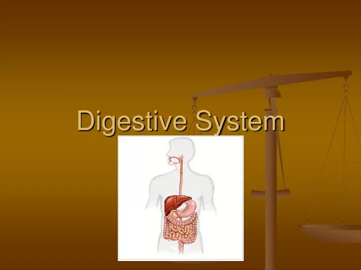
Function • Take in food • Process food - Digestion • Absorb nutrients
Homeostasis of Blood Glucose • What are the two main nutrients carried by blood for cellular respiration? • Why must blood glucose levels be maintained at a certain level?
Nutrition • Animals must supply carbon skeletons necessary to build all needed molecules. • Animals must supply themselves with essential nutrients that can not be made with their enzymes. • Minerals and vitamins
Ingestion – Where is the animal in the Food Web • Herbivores – eat only plants, mouthparts adapted to their food source, coevolution with bacteria and protists to digest cellulose, longer small intestine • Carnivores – eat only animals, mouthparts, shorter small intestine • Omnivores – somewhere in between!
Feeding Mechanisms • Suspension feeding – food particles removed from water • Substrate feeding – live on the food source e.g. leaf miners, dung beetles • Deposit feeding – earthworms take in soil and remove organic material • Fluid feeding – mosquito, leaches • Bulk feeding – large meals, not much chewing
Food Processing • Ingestion – Digestion – Absorption • Ingestion – teeth – dentition, tongue • Digestion physical or chemical – hydrolytic enzymes • Absorption – once food is broken down, the materials must be absorbed and transported throughout the body.
Intracellular Digestion • Paramecium example • paramecium
Gastrovascular Cavity • Hydra and planarian
Alimentary Canal • Mouth EsophagusStomachSmall IntestineLarge IntestineRectumAnus • Mouth digestion of starch via amylase • Stomach – pepsin begins digestion of protein into smaller polypeptides • Small intestine – breakdown and absorb nutrients • Large intestine – reabsorption of water and minerals
Gastrointestinal System • Function-physical and chemical breakdown of food • Includes alimentary canal and accessory organs • Accessory organs: Salivary glands, tongue, teeth, liver, gallbladder, and pancreas
Alimentary canal • Mouth- physical and chemical breakdown. • Mastication-the act of chewing • Saliva contains enzyme amylase to break down carbohydrates
Pharynx- contains opening to trachea as well. Epiglottis covers opening of trachea. • Esophagus-muscular tube dorsal to trachea • Relies on a rhythmic wave-like motion called peristalsis
Sphincter-a circular muscle that constricts a passage or closes a natural orifice (opening) • Cardiac sphincter • Pyloric sphincter • Food sits in stomach for 1-4 hours. Gastric juices contain hydrochloric acid- activates pepsin, kills bacteria.
Small intestine: not so small! 20ft x 1’’ • Duodenum- 1st 10 inches. Bile and pancreatic juice enter here • Jejunum- next 8 ft. • Ileum- final 12 ft. When food leaves small intestine, digestion is complete
Small intestine has lots of enzymes: peptidases • maltase, sucrase, lactase, amylase. • Lipase • Bile Digested food is absorbed into the bloodstream.
Large intestine- 5 ft x 2’’. Separated from small intestine by ileocecal valve • Final absorption of water, storage of indigestible material, absorption of vitamins B and K by bacteria. • Colon connects to rectum- anal canal opens to the anus (final opening). Fecal material is expelled.
But Wait! What about the accessory organs: Liver, gall bladder and pancreas • Liver- largest gland in your body • Secretes bile- emulsifies fat, makes them water soluble. • Stores glucose in the form of glycogen • Makes clotting proteins • Detoxifies blood
Gall bladder- stores and concentrates bile • Pancreas- produces insulin, pancreatic juices amylase and lipase. Insulin regulates the uptake of glucose by the cells
Questions • What parts of the model accurately portrayed the action of the digestive system? • How could we improve this model of digestion? • What organs are play a part in digestion, but are not present in the alimentary canal?
Stomach example • Physical churning • Acid released by cells in response to the hormone Gastrin that is released when a person eats. • Pepsinogin is also released by separate cells – the acid in the stomach changes the shape of pepsinogin to its active form pepsin. Pepsin stimulates more acid – positive feedback. • Acid and Pepsinogin do not mix until they reach the lumen of the stomach or else it would digest the stomach. Stomach lumen is coated with mucus to protect it.
Stomach continued • The food + water + acid and enzymes = acid chyme which passes to the the duodenum of the small intestine. • Ulcers – most caused by a bacteria called Helliobacter pylori treated with antibiotics
Small Intestine • Duodenum – beginning of small intestine the chyme is acidic, digestive enzymes are secreted by the pancreas, liver, and gall bladder (bile), but are active in a neutral pH. Cells add Sodium bicarbonate to neutralize the acid chyme. • Jejunum – villi and micro villi absorb nutrients
The End of the Line • 7 liters of liquid are passed through the stomach and small intestine each day. • Much is reabsorbed by microvilli into lacreal cells in the small intestine. • More water and minerals are reabsorbed by the large intestine. • Food digestion takes 12-24 hours. Longer = less water, Shorter = more water
And then • More cellulose in the diet = faster movement. • E. coli lives in the large intestine on undigested food. The bacteria produce vitamins and some strains help to fight off infections by out-competing the bad guys.
Interesting Adaptations • Herbivores have a longer cecum – usually with symbiotic prokaryotes and protists that digest cellulose. • Rabbits have their symbionts living in their large intestine instead. They eat the digested food (poo) from the first time around and the typical pellets you see are food that has been through twice.
Ruminents • Deer, cows, horses • They have a four chambered alimentary canal. They end up “chewing the cud” cud is made up of food after it has been metabolized by the protists and bacteria. • It passes through the digestive system again. • Ruminents actually get most of their nutrition from the digestion of the microorganisms living inside of them!