Download
1 / 22
220 likes | 401 Views
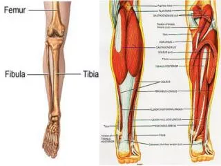
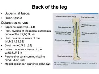
Back of the leg. By Prof. Saeed Abuel Makarem. Muscles of the back of the leg. It is divided into 2 groups: Superficial : 3 muscles , Gastrocnemius. Plantaris. Soleus. Deep: 4 muscles , Popliteus. Flexor digitorum longus. Flexor hallucis longus. Tibialis posterior. Gastrocnemius.

E N D
Back of the leg By Prof. Saeed Abuel Makarem
Muscles of the back of the leg • It is divided into • 2 groups: • Superficial:3 muscles, • Gastrocnemius. • Plantaris. • Soleus. • Deep:4 muscles, • Popliteus. • Flexor digitorum longus. • Flexor hallucis longus. • Tibialis posterior.
Gastrocnemius • Origin: 2 heads • Med: popliteal surface of the femur above medial condyle. • Lat: lateral aspect of lateral condyle of femur. • Insertion: through the tendo calcaneus to post surface of calcaneus. • Nerve: Tibial nerve. • Action: • Flexion of knee joint • Planter flexion of the foot.
Plantaris • Small fusiform muscle. • May be absent or doubled • Homologues to palmaris longus • Origin: lateral supracondylar ridge of the femur. • Insertion: long ribbon- like tendon descends between Gastrocnemius & soleus. • then on medial side of tendo Achillesto t he back of the calcaneus. • Nerve: Tibial nerve. • Action: assists in • Flexion of the knee. • Planter flexion of the foot.
Soleus • It is a broad flat muscle • It forms the main bulk of the calf. • Origin: inverted V-shaped from soleal line of tibia, upper ¼th of back of the fibula & fibrous arch between tibia and fibula. • Insertion: tendo calcaneus • Nerve: Tibial nerve. • Action: • Planter flexion of ankle. • Main forward propulsive force in walking & running with Gastrocnemius & plantaris
Deep Group Popliteus • Origin: lateral surface of lateral condyle of the femur • It is intra capsular, it takes partial origin from the lateral semilunar cartilage. • Insertion: post. surface of the tibia above the soleal line. • Nerve: Tibial nerve. • Action: • Unlocking of the knee joint • Flexion of the knee. • NB. tendon of popliteus separates between lateral meniscus and lateral collateral ligament of the knee
Flexor digitorum longus • Origin:medial part of back of the tibia below the soleal line. • Insertion: terminal phalanges of lateral four toes. • Each slip pierces the tendon of flexor digitorum brevis of the sole. • Nerve: Tibial nerve • Action: • Planter flexion of the terminal phalanx of the lateral 4 toes. • Assists in planter flexion of the foot
Flexor hallucis longus • Origin: lower 2/3rd of the posterior surface of the fibula • Insertion: Base of the distal phalanx of the big toe. • Nerve: Tibial nerve. • Action: Planter flexion of the distal phalanx of big toe • Assists in planter flexion of the foot. • Maintenance of medial longitudinal arch of the foot.
TIBIALIS POSTERIOR • Origin: • Back of interosseous membrane. • Back of tibia lateral to vertical line • Back of fibula medial to medial crest • Insertion: • All tarsus except talus. • The main insertion into tuberosity of the navicular bone. • It is also inserted into the base of 2nd,3rd& 4th metatarsal bones. • Nerve: Tibial nerve • Action: • Planter flexion • Inversion • Maintain the medial longitudinal arch.
Posterior tibial artery • One of the two terminal branches of popliteal artery • Begins at level of the distal border of popliteus muscle • Passes downward deep to Gastrocnemius & soleus • It descends on posterior surface of tibialis posterior • Its lower part lies on back of tibia covered by skin & fascia • It passes behind the medial malleolus to the sole
T D A V N H H Tome: Tibialis posterior Does: Flexor digitorum longus A: Posterior tibial artery V: Posterior tibial vein N: Posterior tibial nerve Hat: Flexor hallucis longus
Branches of posterior tibial artery (PTA): • 1- Peroneal artery: • Arises close to the origin of PTA. • Gives nutrient artery to the fibula & descends behind it. • Gives muscular branches • Shares in anastomosis around the ankle • 2- Muscular branches • 3- Nutrient artery to tibia • 4-Medial planter artery • 5- Lateral planter artery
Tibial Nerve • Larger of the 2 terminal branches of sciatic nerve in the lower 1/3 of back of thigh • It bisects the popliteal fossa • It passes deep to the Gastrocnemius and soleus • It lies on posterior surface of tibialis posterior • It accompanies the posterior tibial artery. • It passes behind the medial malleolus to reach the sole • Branches: • Muscular branches. • Medial calcaneal branch to • Articular to ankle joint • Medial & lateral planter nerves
Sciatic Nerve Injury • The sciatic nerve (L4 and 5 and S1, 2, & 3) curves laterally and downward through the glutealregion. • It leaves the pelvis through the greater sciatic foramen below the piriformis. • situated at first midway between the post. superior iliac spine and the ischial tuberosity. • Then, it lies midway between the tip of the greater trochanter and the ischial tuberosity. • Then it passes downward in the midline on the posterior aspect of the thigh and divides into the common peroneal and tibial nerves, at a variable site above the popliteal fossa. Prof. Saeed Abuel Makarem
Trauma The sciatic nerve is sometimes injured by: • penetrating wounds, • fractures of the pelvis, or • dislocations of the hip joint. Prof. Saeed Abuel Makarem
The sciatic nerve is most frequently injured by…? - badly placed intramuscular injections in the gluteal region. • To avoid this injury, injections into the gluteus maximus or the gluteus medius should be made… • ….well forward on the upper outer quadrant of the buttock. • Most nerve lesions are incomplete, and in 90% of injuries, the common peroneal part of the nerve is the most affected. Why? - The common peroneal nerve fibers lie most superficial in the sciatic nerve. Prof. Saeed Abuel Makarem
The following clinical features are present: Motor: • The hamstring muscles are paralyzed, but weak flexion of the knee is possible. Why? - because of the action of the sartorius(femoral nerve) and gracilis(obturator nerve). • All the muscles below the knee are paralyzed, and the weight of the foot causes it to assume the plantar-flexed position, or foot drop. Prof. Saeed Abuel Makarem
Sensory: • Sensation is lost below the knee,except for a narrow area down the medial side of the lower part of the leg and along the medial border of the foot as far as the ball of the big toe, which is supplied by the saphenous nerve (femoral nerve). Prof. Saeed Abuel Makarem
Sciatica • Sciatica describes the condition in which patients have pain along the sensory distribution of the sciatic nerve. • Thus the pain is experienced in the posterior aspect of the thigh, the posterior and lateral sides of the leg, and the lateral part of the foot. Prof. Saeed Abuel Makarem
Sciatica can be caused by: • prolapse of an intervertebral disc, with pressure on one or more roots of the lower lumbar and sacral spinal nerves, • pressure on the sacral plexus or sciatic nerve by an intrapelvic tumor, or • inflammation of the sciatic nerve or its terminal branches. Prof. Saeed Abuel Makarem