Download
1 / 30
310 likes | 334 Views
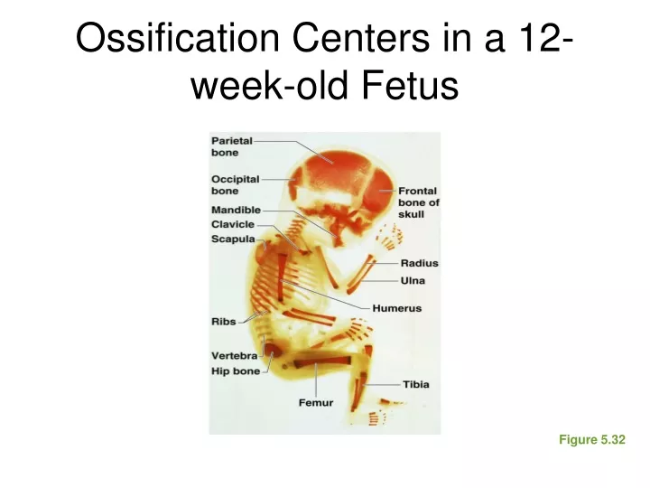
Ossification Centers in a 12-week-old Fetus. Figure 5.32. Types of Bone Cells. Osteocytes—mature bone cells Osteoblasts—bone-forming cells Osteoclasts—bone-destroying cells Break down bone matrix for remodeling and release of calcium in response to parathyroid hormone

E N D
Ossification Centers in a 12-week-old Fetus Figure 5.32
Types of Bone Cells • Osteocytes—mature bone cells • Osteoblasts—bone-forming cells • Osteoclasts—bone-destroying cells • Break down bone matrix for remodeling and release of calcium in response to parathyroid hormone • Bone remodeling is performed by both osteoblasts and osteoclasts
Articularcartilage Hyalinecartilage Spongybone New center ofbone growth New boneforming Epiphysealplatecartilage Growthin bonewidth Medullarycavity Bone startingto replacecartilage Bloodvessels Growthin bonelength New boneforming Bone collar Hyalinecartilagemodel Epiphysealplate cartilage In an embryo In a fetus In a child (a) Figure 5.4a, step 3
Articularcartilage Hyalinecartilage Spongybone New center ofbone growth New boneforming Epiphysealplatecartilage Growthin bonewidth Medullarycavity Bone startingto replacecartilage Bloodvessels Growthin bonelength New boneforming Bone collar Hyalinecartilagemodel Epiphysealplate cartilage In an embryo In a fetus In a child (a) Figure 5.4a, step 3
The Fetal Skull Figure 5.13b
Skeletal Changes Throughout Life • Adolescence • Epiphyseal plates become ossified and long bone growth ends • Size of cranium in relationship to body
Skeletal Changes Throughout Life Figure 5.33a
Bone Fractures • Fracture—break in a bone • Types of bone fractures • Closed (simple) fracture—break that does not penetrate the skin • Open (compound) fracture—broken bone penetrates through the skin • Bone fractures are treated by reduction and immobilization
Repair of Bone Fractures • Hematoma (blood-filled swelling) is formed • Break is splinted by fibrocartilage to form a callus • Fibrocartilage callus is replaced by a bony callus • Bony callus is remodeled to form a permanent patch
Hematoma Hematomaformation Stages in the Healing of a Bone Fracture Figure 5.5, step 1
Hematoma Externalcallus Internalcallus(fibroustissue andcartilage) Newbloodvessels Spongybonetrabecula Hematomaformation Fibrocartilagecallus formation Stages in the Healing of a Bone Fracture Figure 5.5, step 2
Hematoma Externalcallus Bonycallus ofspongybone Internalcallus(fibroustissue andcartilage) Newbloodvessels Spongybonetrabecula Hematomaformation Fibrocartilagecallus formation Bony callusformation Stages in the Healing of a Bone Fracture Figure 5.5, step 3
Hematoma Externalcallus Bonycallus ofspongybone Internalcallus(fibroustissue andcartilage) Newbloodvessels Healedfracture Spongybonetrabecula Bone remodeling Hematomaformation Fibrocartilagecallus formation Bony callusformation Stages in the Healing of a Bone Fracture Figure 5.5, step 4
Inflammatory Conditions Associated with Joints • Bursitis—inflammation of a bursa usually caused by a blow or friction • Arthritis—inflammatory or degenerative diseases of joints • Over 100 different types • The most widespread crippling disease in the United States
Clinical Forms of Arthritis • Osteoarthritis • Most common chronic arthritis • Probably related to normal aging processes • Rheumatoid arthritis • An autoimmune disease—the immune system attacks the joints • Symptoms begin with bilateral inflammation of certain joints • Often leads to deformities
Clinical Forms of Arthritis • Gouty arthritis • Inflammation of joints is caused by a deposition of uric acid crystals from the blood • Can usually be controlled with diet
The Vertebral Column Figure 5.16
Skeletal Changes Throughout Life • Osteoporosis • Bone-thinning disease afflicting • 50% of women over age 65 • 20% of men over age 70 • Disease makes bones fragile and bones can easily fracture • Vertebral collapse results in kyphosis (also known as dowager’s hump) • Estrogen aids in health and normal density of a female skeleton
Skeletal Changes Throughout Life Figure 5.34
Skeletal Changes Throughout Life Figure 5.35