Download
1 / 10
150 likes | 461 Views
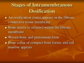
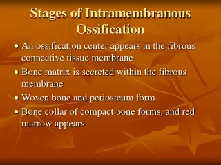
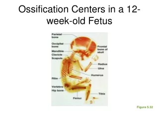
Ossification. Features of Bone. Type one collagen fibres (Think b ONE ) Periosteum is external surface Endosteum lines internal surfaces Mineral component – hydroxyapatite which is made up of calcium and phosphate Rich blood supply. Intramembranous ( i n flat bones).

E N D
Features of Bone • Type one collagen fibres (Think bONE) • Periosteum is external surface • Endosteum lines internal surfaces • Mineral component – hydroxyapatite which is made up of calcium and phosphate • Rich blood supply
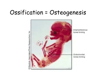
Intramembranous (in flat bones) • Mesenchyme condenses and is highly vascular • Mesenchymal cells differentiate into osteoblasts • Osteoblasts secrete osteoid • Osteoblasts trapped in ECM mature into osteocytes to form woven bone • Bone is reorganised into lamellae bone
Endochondral(at primary and secondary ossification centres) • Chondrocytes in the centre of the cartilage model undergo hypertrophy and secrete ECM • This calcifies and results in chondrocyte death • Osteogenic buds invade dead cartilage, blood vessels bring in osteogenic cells • Osteoblasts develop and secrete osteoid, which becomes mineralised • Woven bone replaces cartilage, perichondrium becomes periosteum, bone reorganised into lamellae bone
Tips for learning this…. • IntraMembranous: starts with Mesenchymal cells. Steps Spell MMOOB • EndoCHONDral: CHONDrocytes. Involves Cartillage
Primary Vs Secondary ossification centres *The bone collar is a cuff of periosteal bone that forms around the diaphysis of the hyaline cartilage model in developing long bones
Fracture Healing – the full version • Haematoma formation: Bleeding from small vessels; periosteal & nutrient arteries • Inflammatory reaction - macrophages invade and remove avascular dead bone around # site • Ingrowth of granulation tissue into the haematoma • Vascular connective tissue / cell proliferation / cytokines & growth factors • Callus formation – bridges gap between bone ends – fibrocellular material initially • Osteogenic cells proliferate from the periosteum • Closer to # site cells differentiate into chondrocytes - islands of cartilage laid down which is soft & radiolucent • Osteogenic cells further from the # site differentiate into osteoblasts & produce woven bone • Cartilaginous components of the callus are gradually replaced by bone through endochondralossification • When the # callus becomes sufficiently firm that movement no longer takes place at the # site it is “clinically united” • However much more needs to be done before the bone is restored to its original strength • Primary callus is gradually replaced by mature lamellar bone • Remodelling by osteoclasts and osteoblasts of the lamellar bone into an appropriate form related to function. Excessive callus resorbed & medullary cavity re-established
Fracture healing: what I would know Haematoma Granulation tissue Callus Woven bone Lamellar bone Remodelling • Haematoma formation • Inflammation reaction. Macrophages invade and remove dead bone • Ingrowth of granulation tissue – vascular tissue • Callus formation – fibrocellular material initially • Osteogenic cells proliferate from the periosteum • Closer to # site cells differentiate into chondrocytes. Osteogenic cells further from the # site differentiate into osteoblasts & produce woven bone • Cartilaginous components of the callus are gradually replaced by bone through endochondralossification. Clinically united • Primary callus is gradually replaced by mature lamellar bone • Remodelling and resorption
Learning Outcomes Explain how bone develops via intramembranous and endochondral ossification Explain the process of bone growth Explain how bones repair following fracture