Download
1 / 12
130 likes | 328 Views
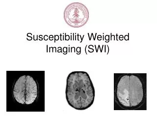
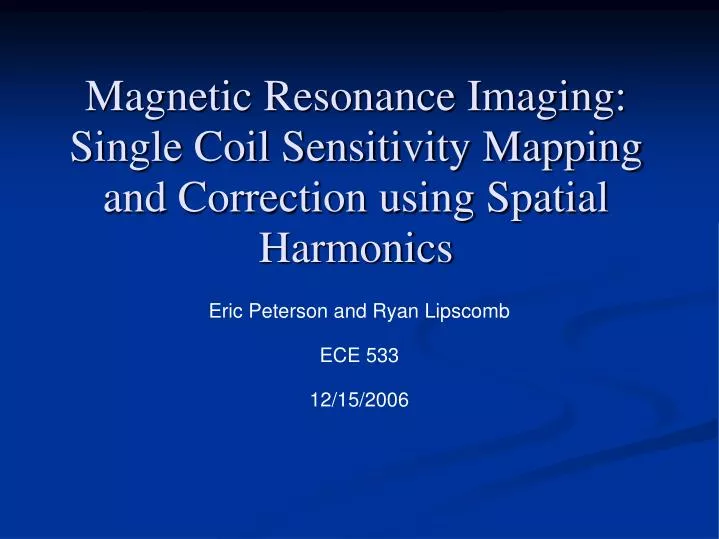
Magnetic Resonance Imaging: Single Coil Sensitivity Mapping and Correction using Spatial Harmonics. Eric Peterson and Ryan Lipscomb ECE 533 12/15/2006. Outline. Motivation Inhomogeneous coil sensitivity Theory Methods Spatial Harmonics Results. Motivation.

E N D
Magnetic Resonance Imaging: Single Coil Sensitivity Mapping and Correction using Spatial Harmonics Eric Peterson and Ryan Lipscomb ECE 533 12/15/2006
Outline • Motivation • Inhomogeneous coil sensitivity • Theory • Methods • Spatial Harmonics • Results
Motivation Flexible Torso Coil from Toshiba Sensitivity profile of a circular phantom using two coils (top and left)
Motivation • Due to the inhomogeneous and varying sensitivity of the vest coil the images vary
Theory Image degradation model Spatial Harmonics equation
Methods • Parallel imaging uses spatial harmonics to define the coil sensitivity map • Use low frequency spatial harmonics to define the sensitivity map • Filter the image and the sensitivity map • Invert the sensitivity map and apply it to the image
Image Input Find noise threshold with a user defined ROI Key Split noise and signal portions of the image Original image Crop Crop Noise only portions of the original image FFT Signal only portions of the original image Select number of harmonics IFFT Sensitivity map Invert and set minimum gain to 1 Inverse sensitivity map Attenuate inverse map using a user defined triangular filter Corrected Image Multiply the inverse map to the image to get the corrected image Normalize the means of the original and corrected images Output Corrected Image with Noise
Results Original Corrected
Results Original Corrected
Results Absolute difference between ADC images before and after correction
Conclusion • This method does a good job of intensity normalization • Preserves high frequency data • The major limitation is due by high frequency components of the lung such as trachea
References • Bydder M, Larkman DJ, Hajnal JV. Generalized SMASH imaging. Magn Reson Med 2002;47:160-170 • Sodickson DK, Manning WJ. Simultaneous acquisition of spatial harmonics (SMASH): fast imaging with radiofrequency coil arrays. Magn Reson Med 1997;38:591-603 • Griswold MA, Jakob PM, Heidemann RM, Nittka M, Jellus V, Wang J, Kiefer B, Haase A. Generalized Autocalibrating Partially Parallel Acquisitions (GRAPPA). Magn Reson Med 2002;47:1202-1210 • Chen XJ, Moller HE, Chawla MS, Cofer GP, Driehuys B, Hedlund LW, Johnson GA. Spatially Resolved Measurements of Hyperpolarized Gas Properties in the Lung In Vivo. Part I: Diffusion Coefficient. Magn Reson Med 1999;42:721–728