Download
1 / 75
750 likes | 769 Views
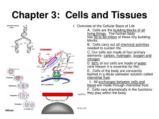
Table of Contents. Chapter: Cells. Section 1: Cell Structure. Section 2: Viewing Cells. Section 3: Viruses. Cell Structure. 1. Common Cell Traits. A cell is the smallest unit that is capable of performing life functions.

E N D
Table of Contents Chapter: Cells Section 1: Cell Structure Section 2: Viewing Cells Section 3: Viruses
Cell Structure 1 Common Cell Traits • A cell is the smallestunit that is capable of performing life functions. • All cells have an outercovering called a cellmembrane. • Inside every cell is a gelatinlike material called cytoplasm. • In the cytoplasm of every cell ishereditarymaterial that controls the life of the cell, DNA.
Cell Structure 1 Comparing Cells • A nerve cell in your leg could be a meter long. • A human egg cell is no bigger than the dot on an i. • A human red blood cell is about one-tenth the size of a human egg cell.
Cell Structure 1 Comparing Cells • A bacterium is even smaller—8,000 of the smallest bacteria can fit inside one of your red blood cells.
Cell Structure 1 Cell Types • Scientists have found that cells can be separated into two groups. • Cells without membrane-bound structures are calledprokaryotic cells.
Cell Structure 1 Cell Types • Cells with membrane-bound structures are called eukaryotic cells.
Cell Structure 1 Sketch and Label! 5 4 3 2 1
Cell Structure 1 Sketch and Label! 5 4 3 2 1
Cell Structure 1 Cell Organization—Cell Wall • The cells of plants, algae, fungi, and most bacteria are enclosed in a cell wall. • Cellwalls are tough, rigid outercoverings that protect the cell and give it shape.
Cell Structure 1 Cell Organization—Cell Wall • A plantcellwallmostly is madeupof a carbohydratecalledcellulose. • Cell walls also can contain pectin, which is used in jam and jelly, and lignin, which is a compound that makescellwallsrigid. • Plant cells responsible for support have a lot of lignin in their walls.
Cell Structure 1 Cell Membrane • The protectivelayeraroundallcellsis the cell membrane. • If cells have cell walls, the cellmembrane is insideofthewall. • The cell membrane regulatesinteractionsbetween the celland the environment.
Cell Structure 1 Cytoplasm • Cells are filledwith a gelatinlike substance called cytoplasm. • Throughout the cytoplasmis a framework called the cytoskeleton, which helps the cellmaintain or change its shape. • The cytoskeleton is made up ofthin, hollowtubes of protein and thin, solid proteinfibers.
Cell Structure 1 Cytoplasm
Cell Structure 1 Cytoplasm • Within the cytoplasm of eukaryotic cells arestructurescalledorganelles. • Some organelles process energy and others manufacture substances needed by the cell or other cells. • Mostorganelles are surroundedbymembranes. • The nucleusis usually the largestorganelle in a cell.
Cell Structure 1 Organelle # 1: Nucleus • The nucleusdirects all cell activities and is separated from the cytoplasm by a membrane. • The nucleuscontains the instructions for everything the cell does.
Cell Structure 1 Nucleus • These instructionsare found on long, threadlike, hereditary material made of DNA. • DNA is the chemical that contains the code for the cell’s structure and activities. • A structure called a nucleolusalso is foundin the nucleus.
Cell Structure 1 Organelle # 2: Chloroplasts • Inplantcells, food is madeingreenorganelles in the cytoplasm calledchloroplasts. • Chloroplastscontain the greenpigmentchlorophyll, which gives many leaves and stems their green color.
Cell Structure 1 Energy-Processing Organelles • These help cells do their work. • Chlorophyllcaptureslightenergy that is used tomake a sugarcalledglucose. • Many cells, including animalcells, donothavechloroplasts for making food. • They must get food from their environment.
Cell Structure 1 Organelle # 3: Mitochondria • The energy in food is stored until it is released by the mitochondria. • Mitochondria (singular, mitochondrion) are organelles whereenergyisreleased from breaking down food into carbon dioxide and water.
Cell Structure • Proteins are partof cell membranes. Other proteins are needed for chemical reactions that take place in the cytoplasm. 1 Organelle # 4: Ribosomes • Cellsmake their own proteinson small structurescalledribosomes.
Cell Structure 1 Organelle # 5: ER • The endoplasmicreticulum, or ER, extends from the nucleus to the cell membrane. • It is a series of foldedmembraneswherematerialsareprocessed and movedaround inside of the cell.
Cell Structure 1 Processing, Transporting, and Storing Organelles • The endoplasmicreticulum may be “rough” or “smooth.” • noattachedribosomesis = smoothER. • roughER= hasribosomes.
Cell Structure 1 Organelle # 6: Golgi • Golgibodiespackage substances.
Cell Structure 1 Organelle # 7: vesicle • The vesiclesdeliver cellular substances to areas inside the cell.
Cell Structure 1 Organelle # 8: vacuole • vacuoles= storage of materials. • A vacuole can store water, waste products, food, and other cellular materials.
Cell Structure 1 Organelle # 9: lysosome • lysosomesbreakdownfood
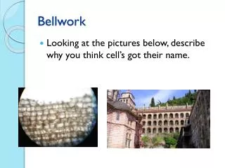
Viewing Cells 2 Cell Theory • In 1665, Robert Hooke cut a thin slice of cork and looked at it under his microscope. • To Hooke, the cork seemed to be made up of empty little boxes, which he named cells.
Viewing Cells 2 Cell Theory • In the 1830s, Matthias Schleiden used a microscope to study plants and concluded that all plants are made of cells. • Theodor Schwann, after observing different animal cells, concluded that all animals are made up of cells.
Viewing Cells 2 Cell Theory • Several years later, Rudolph Virchow hypothesized that cells divide to form new cells. • His observations and conclusions and those of others are summarized in the celltheory.
Viewing Cells 2 Fill in B2 – B4!
Cell Structure 1 From Cell to Organism • A tissue is a groupofsimilarcells that work together to do one job. • Tissues are organizedintoorgans.
Cell Structure 1 From Cell to Organism • An organ is a structure made up of two or more different types of tissues that work together.
Cell Structure • A group of organs working together to perform a certain function is an organ system. Your heart, arteries, veins, and capillaries make up your cardiovascular system. 1 From Cell to Organism Click box to view movie.
Section Check 1 Question 1 Which of these cells is a bacterium? A B
Section Check 1 Answer Prokaryotic cells are only one-celled organisms, suchasbacteria. A
Section Check 1 Question 2 Which part of the cell gives it shape? Answers: A: Nucleus B: Membrane C: Organelle D: Ribosome B
Section Check 1 Question 3 In what part of the cell is the cytoskeleton found? Answers: A: Membrane B: Wall C: Cytoplasm D: Nucleus C
Section Check 1 Question 4 Which cell type LACKS a nucleus? Answers: A: eukaryotic B: plant C: animal D: prokaryotic D
Section Check 1 Question 5 What do ribosomes do? Answers: A: package things B: transport things C: make proteins D: make energy C
Viewing Cells 2 Magnifying Cells • To see most cells, you need to use a microscope. • A microscope has one or more lenses that enlarge the image of an object as though you are walking closer to it.
Viewing Cells 2 Early Microscopes • Made things larger, but not always CLEARER. • In the mid 1600s, Antoine vanLeeuwenhoek, a Dutch fabric merchant, made a simple microscope with a tiny glass bead for a lens.
Viewing Cells 2 Modern Microscopes • Use lenses to bend light. • Can be simple or compound. • Stereo: more of a 3-D image. • Powers multiplied together =totalmagnification..
Viewing Cells 2 Electron Microscopes • Things that are too small to be seen with other microscopes can be viewed with an electron microscope. • Instead of using lenses to direct beams of light, an electron microscope usesa magnetic field in a vacuum to direct beams of electrons.
Viewing Cells 2 Electron Microscopes • Scanning electron microscopes (SEM) produce a realistic, three-dimensional image. • Only the surface of the specimen can be observed using an SEM.
Viewing Cells 2 Electron Microscopes • Transmission electron microscopes (TEM) produce a two-dimensional image of a thinly-sliced specimen. • Results must be photographed or captured in other ways.
Section Check 2 Question 1 Who developed a microscope using a tiny glass bead for a lens? A. Antoine van Leeuwenhoek B. Edward Jenner C. Matthias Schleiden D. Theodor Schwann
Section Check 2 Answer A Antoine van Leeuwenhoek
Section Check 2 Question 2 How many lenses does a simple microscope have? A. 0 B. 1 C. 2 D. 4
Section Check 2 Answer B - 1