Download
1 / 23
230 likes | 368 Views
Chapter 1 Cells: discovery and exploration. Cells are the basic function units of all living things. Most cells are too small to be seen by the naked eye, so microscopes give enlarged images of cells and the structures they contain. They make it possible to examine cells in great detail.

E N D
Cells are the basic function units of all living things. Most cells are too small to be seen by the naked eye, so microscopes give enlarged images of cells and the structures they contain. They make it possible to examine cells in great detail.
The recognition that all living things share a common structural unit – the cell – provided the foundation for one of the major unifying themes in biology. • But…how are cells made?
Scientists Schwann and Schlieder came up with the first main theory, known as the Cell Theory. All living things consist of one or more organised structures that are called cells or of products of cells. Cells are the basic functional unit of life.
Several ideas were put forward as to how cells became cells. Spontaneous generation is the idea that living things could arise from non-living or dead matter. Another idea was that living things developed that gathered to form a compact mass, then becoming organised cells.
Life span of cells The lifespan of cells in multi-cellular organisms varies greatly, even among human cells The average life span of some human cells are: • Stomach cells 2 days • Mature sperm cells 2-3 days • Skin cells 20-35 days • Red blood cells about 120 days
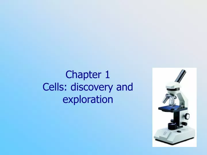
Tools for viewing cells Generally, cells are too small to be seen with the naked eye. Because of this, we need to use microscopes. Microscopes can be divided into two main categories: • Light microscopes and • Electron microscopes
Light microscopes (LMs) • Increase the ability of the human eye to see tiny objects • Use visible light that illuminates and passes through a specimen
Simple light microscopes are similar to a magnifying glass. Compound light microscopes have at least two sets of lenses. Most have several objective lenses, each of a different magnification. How large the object appears depends on the magnification powers of both the eyepiece and the lenses used.
Characteristics of the lenses also influence a microscope’s resolution. Resolution is the ability to see two points that are close together as two separate points. We use microscopes to resolve things that our eyes are unable to see.
Staining Cells are virtually colourless, so are difficult to see under a standard LM. Therefore, staining is usually required. Groups of cells are cut into thin slices, only a few cells thick before staining. These treatments are often toxic to the cells, and can often distort cell features.
Other LMs • Phase contrast LM (see Fig 1.11b) • A modified compound light microscope (CLM) • Developed to view unstained living cells • The image developed has highly contrasting light and dark areas • Flourescence microscope (see Fig 1.12) • Another type of CLM • Uses UV light to reveal compounds that have been stained with flourescent dyes e.g. cancerous breast cells
Other LMs • Scanning confocal microscope (see Fig 1.14) • Sort of a cross between a light microscope and an electron microscope • Uses laser light and special optics to allow a viewer to look at successively deeper layers of an object • The viewer does not have to cut the object into thin sections, the microscope does that • Can also produce 3D
Electron Microscopes • Transmission electron microscopes (TEM) (see Fig 1.18) • Invented in the 1930’s • A beam of electrons with a much shorter wavelength passes through and is used to illustrate specimens • The beam is shone through the specimen, revealing great detail of the internal structure of specimens
TEMs have much greater resolving power than light microscopes. This is because of the shorter wavelengths of electron beams. TEMs have revealed the presence of many kinds of cell organelles and have shown the internal structure that exists within cells.
Scanning electron microscope (SEM) (see Fig 1.19) • Released in 1965 • The electrons are bounced off the surface of the specimen, giving an extremely detailed view of the surface of a specimen. • Depending on the size, parts of or whole organisms can be scanned
Which type of electron microscope was used to generate these images?
Recent developments - LMs • Differential interference contrast (DIC) microscopes • Used to obtain 3D impressions of an object • Used in IVF techniques to view the process of the sperm fertilising the egg (see FIG 1.22) • Automatic scanning of cells - Used to search many cells at a time for defective cells
Recent developments - EMs • Freeze fracture • A small block of living or dead cells are rapidly frozen in liquid nitrogen • Such rapid freezing reduces changes to the cells, so that when they are put into the vacuum chamber to be cut, the internal structures can easily be seen • Shadowing (see Fig 1.24) • Fractured pieces of cells are exposed to and covered by heavy metals e.g. platinum or gold • Specimens are then dissolved away, and the metals remain with the impressions of the cells distinguishable features
Images were taken from… http://www.microscopeworld.com/misc/gift-ideas-131.htm http://bomi.ou.edu/bot1114/botany1114/elder/cells/acell2.gif http://sun.menloschool.org/~cweaver/cells/plantcell /url?q=http://www.seallabs. com/graphics/ant.jpg www.lps.u-psud.fr/.../ Image/Materiel/Orsay1.jpg http://www.cas.muohio.edu/~mbi-ws/microscopes/images/LightDiagram.GIF http://www.columbia.edu/cu/record/archives/vol21/vol21_iss6/record2106.32c.gif http://history.nih.gov/exhibits/genetics/images/sect2/9b.jpg Reference for text… Kinnear J and Martin M. 2006. Nature of Biology, Book 1, Third Edition. Jacaranda, Q.L.D.