Download
1 / 30
330 likes | 392 Views
Spirochaetales Treponema, Borrelia, Leptospira Dr. Hala Al Daghistani. Taxonomy. Order : Spirochaetales Family : Spirochaetaceae Genus : Treponema Borrelia Family : Leptospiraceae Genus : Leptospira. Gram-negative spirochetes Spirochete from Greek for “coiled hair”

E N D
Spirochaetales Treponema, Borrelia, Leptospira Dr. Hala Al Daghistani
Taxonomy • Order: Spirochaetales • Family: Spirochaetaceae • Genus: Treponema • Borrelia • Family: Leptospiraceae • Genus: Leptospira
Gram-negative spirochetes • Spirochete from Greek for “coiled hair” • Extremely thin and can be very long • Motile by periplasmic flagella (axial fibrils or endoflagella) • Outer sheath encloses axial fibrils • Axial fibrils originate from insertion pores at both poles of cell
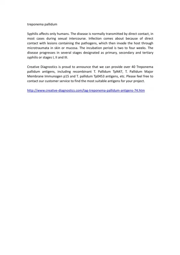
Venereal Treponemal Disease • - T. pallidum are slender spirals with spiral coils spaced at a distance of 1 μm from one another. • - Pathogenic T pallidum has never been cultured on artificial media, in fertile eggs, or in tissue culture. • - Nonpathogenic treponemes (Reiter strain) can be cultured anaerobically in vitro. • T pallidum is a microaerophilic organism; it survives best in 3–5% oxygen. • Cause Syphilis • Primarily sexually transmitted disease • May be transmitted congenitally
General Characteristics of Treponema pallidum • Too thin to be seen with light microscopy in specimens stained with Gram stain or Giemsa stain • Motile spirochetes can be seen with darkfield micoscopy • Staining with anti-treponemal antibodies labeled with fluorescent dyes • Intracellular pathogen • Cannot be grown in cell-free cultures in vitro • Koch’s Postulates have not been met • Do not survive well outside of host
Epidemiology of T. pallidum • Transmitted from direct sexual contact or from mother to fetus • Long incubation period during which time host is non-infectious
Virulence Factors of T. pallidum • Outer membrane proteins promote adherence • Hyaluronidase may facilitate perivascular infiltration • Antiphagocytic coating of fibronectin • Tissue destruction and lesions are primarily result of host’s immune response (immunopathology)
Pathogenesis of T. pallidum • Primary disease • Spirochetes multiply locally at the site of entry, and some spread to nearby lymph nodes and then reach the bloodstream. • A papule develops at the site of infection and breaks down to form an ulcer with a clean, hard base ("hard chancre“)
This "primary lesion" always heals spontaneously, but 2–10 weeks later the "secondary" lesions appear. These consist of a red maculopapular rash anywhere on the body, including the hands and feet, anogenital region, axillas, and mouth. There may also be syphilitic meningitis, chorioretinitis, hepatitis, nephritis.
Latent Stage Syphilis & Tertiary • In Latent syphilis, which can last for years, there are little to no symptoms. • First 4 years = Early latent • Subsequent period = Late latent • In Tertiary syphilis there are Gummas (soft swelling, granulomatous lesions) which occurs in the connective tissue of the liver, brain, testes, and heart, skin, bones, and liver; degenerative changes in the central nervous system (meningovascular syphilis); or cardiovascular
Congenital Syphilis • Congenital syphilis results from transplacental infection • T. pallidumsepticemia in the developing fetus and widespread dissemination • Some of the infected fetuses die, miscarriages result; others are stillborn at term. • Others are born live but develop the signs of congenital syphiliswith variety of central nervous system anomalies.
Cardiolipinis an important component of the treponemal antigens. the spirochetes cause the development of antibody-like substance, reagin, which gives positive flocculation tests
Giemsa Stain of Borrelia recurrentis in Blood Phase Contrast Microscopy Light Microscopy
Epidemiology of Borrelia Infections Pediculus humanus Borrelia recurrentis Ornithodoros spp. Borrelia spp. Ixodes spp. Borrelia burgdorferi
Borrelia species & relapsing feverRelapsing fever (epidemic) is caused by Borreliarecurrentis, transmitted by the human body louseAntigenic structureThe antigenic structure of the organisms changes in the course of a single infection. Ultimate recovery (after 3 to 10 relapses) is associated with the presence of antibodies against several antigenic variants.
Pathologyfatal cases show spirochetes in great numbers in the spleen and liver, necrotic and hemorrhagic lesions in the kidneys, gastrointestinal tract, spinal fluid and brain of persons who have had meningitis.Pathogenesis & Clinical Findings- The incubation period is 3–10 days - The onset is sudden, with chills and an abrupt rise of temperature.During this time, spirochetes found in the blood. The fever persists for 3–5 days and then declines, leaving the patient weak but not ill.- Afebrile period lasts 4–10 days and is followed by a second attack of chills, fever, intense headache, and malaise. - during the febrile stages, organisms are present in the blood; during the afebrile periods, they are absent.
Borreliaburgdorferi & Lyme Disease Lyme disease is named after the town of Lyme, where clusters of cases in children were identified. Lyme disease is caused by the spirochete B. burgdorferi and is transmitted to humans by the bite of a small ixodes tick. The most common sign of infection is an expanding area of redness on the skin, known as erythemamigrans, that begins at the site of a tick bite about a week after it has occurred. In addition, flu-like symptoms, and late manifestations often with arthralgia and arthritis.
B burgdorferi is a spiral organism, highly motile.Transmission of B burgdorferi to humans is by injection of the organism in tick saliva or by regurgitation of the tick's midgut contents. After injection by the tick, the organism migrates out from the site, producing the characteristic skin lesion. Dissemination occurs by lymphatics or blood to other skin and musculoskeletal sites and to many other organs.A unique skin lesion that begins 3 days to 4 weeks after a tick bite often marks
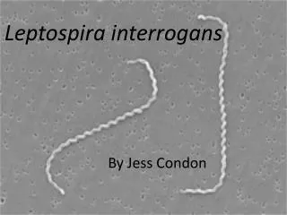
Leptospirosisis an infection caused by corkscrew-shaped bacteria called Leptospira interrogansSigns and symptoms can range from none to mild such as headaches, muscle pains, and fever; to severe with bleeding from the lungs or meningitis.