Download

1 / 107
1.08k likes | 1.12k Views
Spirochaetales ~~~~~~~~~~~~~~~~~~ Treponema Borrelia & Leptospira. Taxonomy. Order : Spirochaetales Family : Spirochaetaceae Genus : Treponema Borrelia Family : Leptospiraceae Genus : Leptospira. General Overview of Spirochaetales. Gram-negative spirochetes

E N D
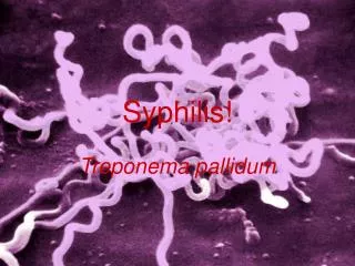
Spirochaetales ~~~~~~~~~~~~~~~~~~ Treponema Borrelia & Leptospira
Taxonomy • Order: Spirochaetales • Family: Spirochaetaceae • Genus: Treponema • Borrelia • Family: Leptospiraceae • Genus: Leptospira
General Overview of Spirochaetales • Gram-negative spirochetes • Spirochete from Greek for “coiled hair” • Extremely thin and can be very long • Tightly coiled helical cells with tapered ends • Motile by periplasmic flagella (a.k.a., axial fibrils or endoflagella) • Outer sheath encloses axial fibrils wrapped around protoplasmic cylinder • Axial fibrils originate from insertion pores at both poles of cell • May overlap at center of cell in Treponema and Borrelia, but not in Leptospira • Differering numbers of endoflagella according to genus & species
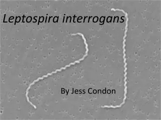
OS = outer sheath AF = axial fibrils Leptospira interrogans AF Tightly Coiled Spirochete
Cross-Section of Spirochete with Periplasmic Flagella Cross section of Borrelia burgdorferi NOTE: a.k.a., endoflagella, axial fibrils or axial filaments. (Outer sheath)
Nonvenereal Treponemal Diseases • Bejel, Yaws & Pinta • Primitive tropical and subtropical regions • Primarily in impoverished children
Treponema pallidum ssp. endemicum • Bejel (a.k.a. endemic syphilis) • Initial lesions: nondescriptoral lesions • Secondary lesions: oral papules and mucosal patches • Late: gummas (granulomas) of skin, bones & nasopharynx • Transmitted person-to-person by contaminated eating utensils • Primitive tropical/subtropical areas (Africa, Asia & Australia)
Treponema pallidum ssp. pertenue (May also see T. pertenue) • Yaws: granulomatous disease • Early: skin lesions (see below) • Late: destructive lesions of skin, lymph nodes & bones • Transmitted by direct contact with lesions containing abundant spirochetes • Primitive tropical areas (S. America, Central Africa, SE Asia) Papillomatous Lesions of Yaws: painless nodules widely distributed over body with abundant contagious spirochetes.
Treponema carateum • Pinta: primarily restricted to skin • 1-3 week incubation period • Initial lesions: smallpruriticpapules • Secondary: enlarged plaques persist for months to years • Late: disseminated, recurrenthypopigmentation or depigmentation of skin lesions; scarring & disfigurement • Transmitted by direct contact with skin lesions • Primitive tropical areas (Mexico, Central & South America) Hypopigmented Skin Lesions of Pinta:depigmentation is commonly seen as a late sequel with all treponemal diseases
Venereal Treponemal Disease • Syphilis • Primarily sexually transmitted disease (STD) • May be transmitted congenitally
General Characteristics of Treponema pallidum • Too thin to be seen with light microscopy in specimens stained with Gram stain or Giemsa stain • Motile spirochetes can be seen with darkfield micoscopy • Staining with anti-treponemal antibodies labeled with fluorescent dyes • Intracellular pathogen • Cannot be grown in cell-free cultures in vitro • Koch’s Postulates have not been met • Do not survive well outside of host • Care must be taken with clinical specimens for laboratory culture or testing
Epidemiology of T. pallidum • Transmitted from direct sexual contact or from mother to fetus • Not highly contagious (~30% chance of acquiring disease after single exposure to infected partner) but transmission rate dependent upon stage of disease • Long incubation period during which time host is non-infectious • Useful epidemiologically for contact tracing and administration of preventative therapy • Prostitution for drugs or for money to purchase drugs remains central epidemiologic aspect of transmission
Pathogenesis of T. pallidum • Tissue destruction and lesions are primarily a consequence of patient’s immune response • Syphilis is a disease of blood vessels and of the perivascular areas • In spite of a vigorous host immune response the organisms are capable of persisting for decades • Infection is neither fully controlled nor eradicated • In early stages, there is an inhibition of cell-mediated immunity • Inhibition of CMI abates in late stages of disease, hence late lesions tend to be localized
Virulence Factors of T. pallidum • Outer membrane proteins promote adherence • Hyaluronidase may facilitate perivascular infiltration • Antiphagocytic coating of fibronectin • Tissue destruction and lesions are primarily result of host’s immune response (immunopathology)
Pathogenesis of T. pallidum (cont.) Primary Syphilis • Primary disease process involves invasion of mucus membranes, rapid multiplication & wide dissemination through perivascular lymphatics and systemic circulation • Occurs prior to development of the primary lesion • 10-90 days (usually 3-4 weeks) after initial contact the host mounts an inflammatory response at the site of inoculation resulting in the hallmark syphilitic lesion, called the chancre (usually painless) • Chancre changes from hard to ulcerative with profuse shedding of spirochetes • Swelling of capillary walls & regional lymph nodes w/ draining • Primary lesion heals spontaneously by fibrotic walling-off within two months, leading to false sense of relief
Pathogenesis of T. pallidum (cont.) Secondary Syphilis • Secondary disease 2-10 weeks after primary lesion • Widely disseminated mucocutaneous rash • Secondary lesions of the skin and mucus membranes are highly contagious • Generalized immunological response
Pathogenesis of T. pallidum (cont.) Latent Stage Syphilis • Following secondary disease, host enters latent period • First 4 years = early latent • Subsequent period = late latent • About 40% of late latent patients progress to late tertiary syphilitic disease
Pathogenesis of T. pallidum (cont.) Tertiary Syphilis • Tertiary syphilis characterized by localized granulomatous dermal lesions (gummas) in which few organisms are present • Granulomas reflect containment by the immunologic reaction of the host to chronic infection • Late neurosyphilis develops in about 1/6 untreated cases, usually more than 5 years after initial infection • Central nervous system and spinal cord involvement • Dementia, seizures, wasting, etc. • Cardiovascular involvement appears 10-40 years after initial infection with resulting myocardial insufficiency and death
Diagram of a Granuloma (a.k.a. gumma in skin or soft tissue) NOTE:ultimately a fibrin layer develops around granuloma, further “walling off” the lesion
Progression of Untreated Syphilis Late benign Gummas in skin and soft tissues Tertiary Stage
Pathogenesis of T. pallidum (cont.) Congenital Syphilis • Congenital syphilis results from transplacental infection • T. pallidumsepticemia in the developing fetus and widespread dissemination • Abortion, neonatal mortality, and late mental or physical problems resulting from scars from the active disease and progression of the active disease state
Comparison of Incidence of 1o & 2o Syphilis in Women and Congenital Syphilis
Prevention & Treatment of Syphilis • Penicillin remains drug of choice • WHO monitors treatment recommendations • 7-10 days continuously for early stage • At least 21 days continuously beyond the early stage • Prevention with barrier methods (e.g., condoms) • Prophylactic treatment of contacts identified through epidemiological tracing
Diagnostic Tests for Syphilis (Original Wasserman Test) NOTE: Treponemal antigen tests indicate experience with a treponemal infection, but cross-react with antigens other than T. pallidum ssp. pallidum. Since pinta and yaws are rare in USA, positive treponemal antigen tests are usually indicative of syphilitic infection.
Review Handout on Sensitivity & Specificity of Diagnostic Tests
Conditions Associated with False Positive Serological Tests for Syphillis
Effect of Treatment for Syphillis on Rapid Plasma Reagin Test Reactivity
Giemsa Stain of Borrelia recurrentis in Blood Phase Contrast Microscopy Light Microscopy
Epidemiology of Borrelia Infections Pediculus humanus Borrelia recurrentis Ornithodoros spp. Borrelia spp. Ixodes spp. Borrelia burgdorferi
Epidemiology of Relapsing Fever • Associated with poverty, crowding, and warfare • Arthropod vectors • Louse-borne borreliosis = Epidemic Relapsing Fever • Transmitted person-to-person by human body lice (vectors) from infected human reservoir • Infect host only when louse is injured, e.g., during scratching • Therefore, a single louse can only infect a single person • Lice leave host that develops a fever and seek normal temperature host • Tick-borne borreliosis = Endemic Relapsing Fever • Sporadic cases • Transmitted by soft body ticks (vectors) from small mammal reservoir • Ticks can multiply and infect new human hosts
Pathogenesis of Relapsing Fever • Relapsing fever (a.k.a., tick fever, borreliosis, famine fever) • Acute infection with 2-14 day (~ 6 day) incubation period • Followed by recurring febrile episodes • Constant spirochaetemia that worsens during febrile stages • Epidemic Relapsing Fever = Louse-borne borreliosis • Borrelia recurrentis • Endemic Relapsing Fever = Tick-borne borreliosis • Borrelia spp.
Pathogenesis of Lyme Borreliosis • Lyme disease characterized by three stages: • Initially a unique skin lesion (erythema chronicum migrans (ECM)) with general malaise • ECM not seen in all infected hosts • ECMoftendescribed as bullseye rash • Lesions periodically reoccur • Subsequent stage seen in 5-15% of patients with neurological or cardiac involvement • Third stage involves migrating episodes of non-destructive, but painful arthritis • Acute illness treated with phenoxymethylpenicillin or tetracycline
Erythema chronicum migrans of Lyme Borreliosis Bullseye rash